. 2020 Jul 20;12(7):463.
doi: 10.3390/toxins12070463.

Effects of Deoxynivalenol and Zearalenone on the Histology and Ultrastructure of Pig Liver

Affiliations

Effects of Deoxynivalenol and Zearalenone on the Histology and Ultrastructure of Pig Liver

Natalia Skiepko et al. Toxins (Basel). .

Abstract

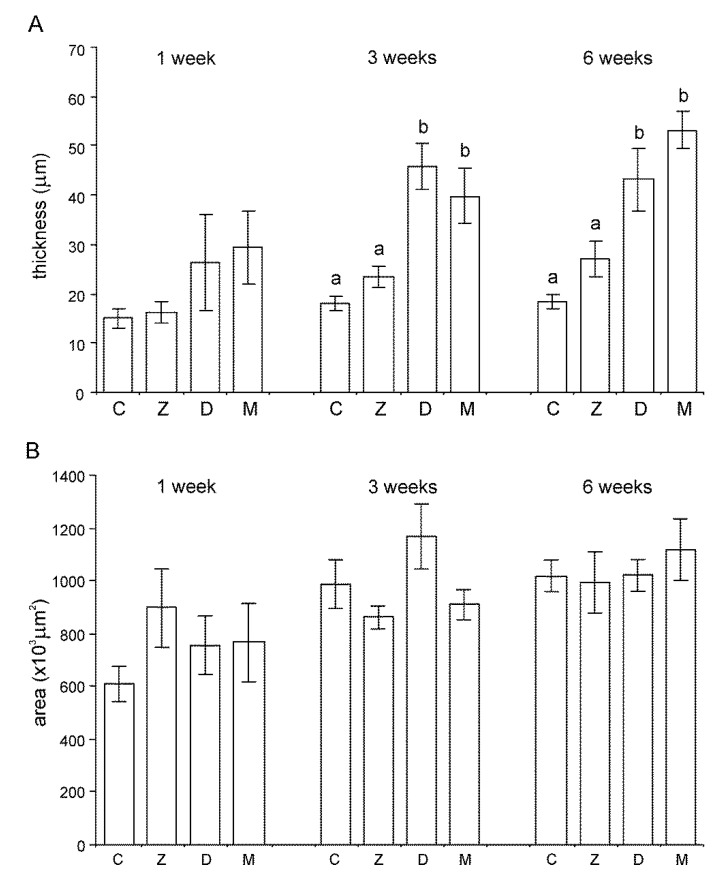
The purpose of this study was to determine the effects of single and combined administrations of deoxynivalenol (DON) and zearalenone (ZEN) on the histology and ultrastructure of pig liver. The study was performed on immature gilts, which were divided into four equal groups. Animals in the experimental groups received DON at a dose of 12 μg/kg body weight (BW) per day, ZEN at 40 μg/kg BW per day, or a mixture of DON (12 μg/kg BW per day) and ZEN (40 μg/kg BW). The control group received vehicle. The animals were killed after 1, 3, and 6 weeks of experiment. Treatment with mycotoxins resulted in several changes in liver histology and ultrastructure, including: (1) an increase in the thickness of the perilobular connective tissue and its penetration to the lobules in gilts receiving DON and DON + ZEN; (2) an increase in the total microscopic liver score (histology activity index (HAI)) in pigs receiving DON and DON + ZEN; (3) dilatation of hepatic sinusoids in pigs receiving ZEN, DON and DON + ZEN; (4) temporary changes in glycogen content in all experimental groups; (5) an increase in iron accumulation in the hepatocytes of gilts treated with ZEN and DON + ZEN; (6) changes in endoplasmic reticulum organization in the hepatocytes of pigs receiving toxins; (7) changes in morphology of Browicz-Kupffer cells after treatment with ZEN, DON, and DON + ZEN. The results show that low doses of mycotoxins used in the present study, even when applied for a short period, affected liver morphology.

Keywords: deoxynivalenol; hepatocyte; histology; liver; mycotoxins; pig; ultrastructure; zearalenone.


Conflict of interest statement

The authors declare no conflict of interest.

Figures

Figure 1
Figure 1
(A,B) Architecture of the liver in a control pig, 6th week of experiment (A) and in a pig receiving deoxynivalenol (DON) for 6 weeks (B). Note the thickening of the interlobular septa. (C) A strip of connective tissue in the liver lobule. A pig was treated with DON for 3 weeks. (D) Focal fibrosis inside the liver lobule of a pig treated with DON for 6 weeks. Note that the central vein is surrounded by connective tissue. Figures (AD) show hematoxylin and eosin stained sections.
Figure 2
Figure 2
(A,B) Trichrome staining of the liver in a control pig, 6th week of experiment (A) and in a pig receiving DON for 6 weeks (B). Pay attention to the thickness of the interlobular septa and the content of collagen fibers in them.
Figure 3
Figure 3
(A) Thickness of the connective tissue septa. (B) Cross-sectional area of the liver lobules. C—control group, Z—group treated with zearalenone (ZEN), D—group treated with deoxynivalenol (DON), and M—group treated with DON and ZEN. The values are presented as mean ± standard deviation. Bars labeled with different small lower-case letters differ significantly at p ≤ 0.05.
Figure 4
Figure 4
Histopathological score of the examined livers according to the histology activity index (HAI) modified by Stanek et al. [38]. Six histopathological parameters were scored in hematoxylin and eosin (HE)-stained tissues (see Figure S1 in Supplementary material), and the cumulative HAI score was calculated for each experimental group. The mean value of the cumulative HAI scores in each group is represented by the total height of the bar. C—control group, Z—group treated with zearalenone (ZEN), D—group treated with deoxynivalenol (DON), and M—group treated with DON and ZEN. The values of the cumulative HAI score labeled with different lower-case letters above the bars differ significantly at p ≤ 0.05.
Figure 5
Figure 5
(A,B) Distribution of glycogen in hepatocytes in a control pig (A) and a pig treated with ZEN + DON (B). The first week of experiment. Periodic acid-Schiff reaction. Note the increase in the amount of glycogen in a pig treated with mycotoxins.
Figure 6
Figure 6
(A,B) Iron deposits in the liver of pigs receiving ZEN + DON for six weeks. (A) Iron deposits in hepatocytes in the form of fine granules. (B) Iron deposits with variable sizes in perilobular connective tissue. Prussian blue staining.
Figure 7
Figure 7
(AF) Ultrastructure of hepatocytes in control pigs, 6th week of the experiment (A,B), pigs treated with ZEN for 6 weeks (C,D) and pigs treated with DON for 6 weeks (E,F). Note the specific organization of endoplasmic reticulum with the predominance of smooth reticulum over rough reticulum in pigs treated with ZEN. In DON-treated animals, both types of endoplasmic reticulum were formed by abundant short, dilated cisterns and vesicles. SD, space of Disse; BC, bile canaliculus; N, nucleus; M, mitochondria; RER, rough endoplasmic reticulum; SER, smooth endoplasmic reticulum; G, glycogen particles.
Figure 8
Figure 8
(A,B) Browicz–Kupffer cells (BK) in the sinusoid of pigs treated with DON for 3 weeks (A) and 6 weeks (B). Note the presence of numerous processes in Figure (A) and lysosomes and rest bodies in Figure (B,C). BK and pit (P) cells in the sinusoid of a pig treated with DON + ZEN for 6 weeks. (D) Ito cell (It) in the perivascular space of pigs treated with DON + ZEN for 6 weeks. Note the cisterns of the rough endoplasmic reticulum.
Figure 9
Figure 9
(A) Accumulation of collagen fiber in the space of Disse in a pig receiving DON for 6 weeks. (B) Penetration of collagen fibers between hepatocytes in a pig treated with DON + ZEN for 6 weeks.

References

    1. Cavret C., Lecoeur S. Fusariotoxin transfer in animals. Food Chem. Toxicol. 2006;44:444–453. doi: 10.1016/j.fct.2005.08.021. - DOI - PubMed
    1. Buszewska-Forajta M. Mycotoxins, invisible danger of feedstuff with toxic effect on animals. Toxicon. 2020;182:34–53. doi: 10.1016/j.toxicon.2020.04.101. - DOI - PubMed
    1. Nayakwadi S., Ramu R., Kumar Sharma A., Kumar Gupta V., Rajukumar K., Kumar V., Shirahatti P.S., Rashmi L., Basalingappa K.M. Toxicopathological studies on the effects of T-2 mycotoxin and their interaction in juvenile goats. PLoS ONE. 2020;15:e229463. doi: 10.1371/journal.pone.0229463. - DOI - PMC - PubMed
    1. Koselski M., Dziubińska H., Trębacz K., Sieprawska A., Filek M. The Role of SV Ion Channels under the Stress of Mycotoxins Induced in Wheat Cells—Protective Action of Selenium Ions. J. Plant Growth Regul. 2019;38:1255–1259. doi: 10.1007/s00344-019-09930-z. - DOI
    1. Sun Y., Wen J., Chen R., Deng Y. Variable protein homeostasis in housekeeping and non-housekeeping pathways under mycotoxins stress. Sci. Rep. 2019;9:7819. doi: 10.1038/s41598-019-44305-0. - DOI - PMC - PubMed

Publication types