. 2020 Jul 20;12(7):1978.
doi: 10.3390/cancers12071978.

Depletion of Macrophages Improves Therapeutic Response to Gemcitabine in Murine Pancreas Cancer

Affiliations

Depletion of Macrophages Improves Therapeutic Response to Gemcitabine in Murine Pancreas Cancer

Soeren M Buchholz et al. Cancers (Basel). .

Abstract

Background: The tumor microenvironment (TME) is composed of fibro-inflammatory cells and extracellular matrix (ECM) components. However, the exact contribution of the various TME compartments towards therapeutic response is unknown. Here, we aim to dissect the specific contribution of tumor-associated macrophages (TAMs) towards drug delivery and response in pancreatic ductal adenocarcinoma (PDAC).

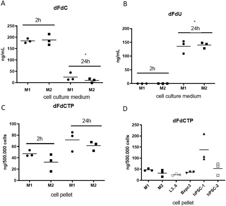
Methods: The effect of gemcitabine was assessed in human and murine macrophages, human pancreatic stellate cells (hPSCs), and tumor cells (L3.6pl, BxPC3 and KPC) in vitro. The drug metabolism of gemcitabine was analyzed by liquid chromatography-tandem mass spectrometry (LC-MS/MS). Preclinical studies were conducted using KrasG12D;p48-Cre and KrasG12D;p53172H;Pdx-Cre mice to investigate gemcitabine delivery at different stages of tumor progression and upon pharmacological TAM depletion.

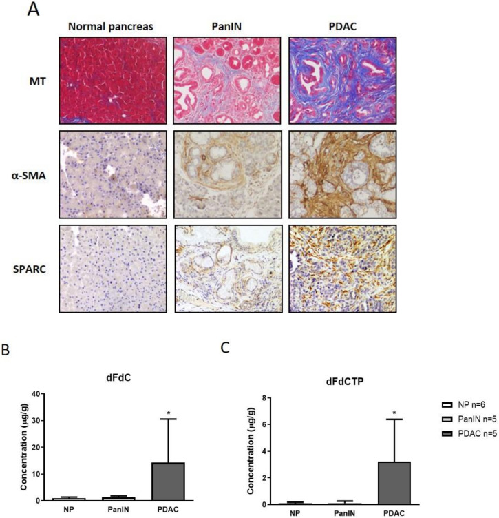
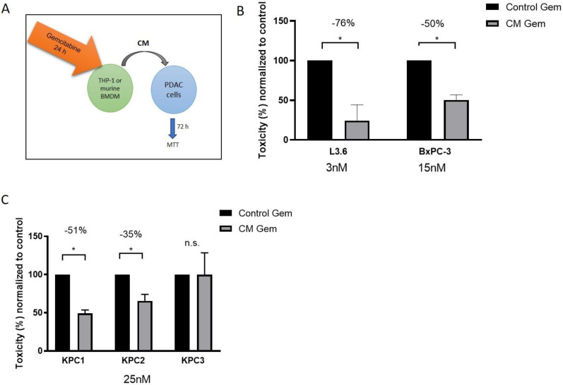
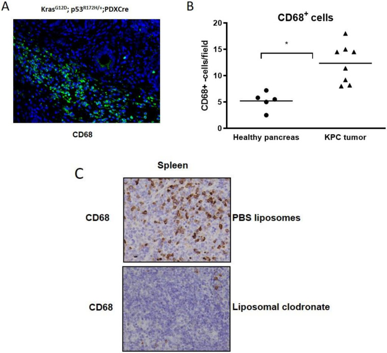
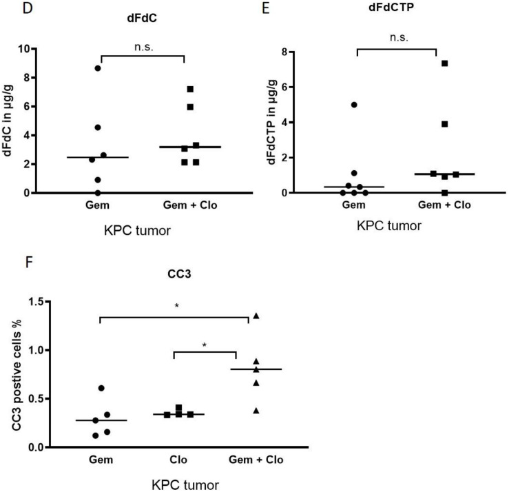
Results: Gemcitabine accumulation was significantly increased in murine PDAC tissue compared to pancreatic intraepithelial neoplasia (PanIN) lesions and healthy control pancreas tissue. In vitro, macrophages accumulated and rapidly metabolized gemcitabine resulting in a significant drug scavenging effect for gemcitabine. Finally, pharmacological TAM depletion enhanced therapeutic response to gemcitabine in tumor-bearing KPC mice.

Conclusion: Macrophages rapidly metabolize gemcitabine in vitro, and pharmacological depletion improves the therapeutic response to gemcitabine in vivo. Our study supports the notion that TAMs might be a promising therapeutic target in PDAC.

Keywords: chemoresistance; drug delivery; macrophages; pancreatic cancer.


Conflict of interest statement

The authors declare no conflict of interest. The funders had no role in the design of the study; in the collection, analyses, or interpretation of data; in the writing of the manuscript, or in the decision to publish the results.

Figures

Figure 1
Figure 1
(A) Representative Masson trichrome (MT) and immunohistochemical stainings for alpha-smooth muscle actin (α-SMA) and secreted protein acidic and rich in cysteine (SPARC) in normal murine pancreas, murine pancreatic intraepithelial neoplasia (PanIN) and pancreatic ductal adenocarcinoma (PDAC) tissues from LSL-KrasG12D/+; p48-Cre (KC) mice showing progressive desmoplastic features. (B,C) KC mice and control B6 mice were treated with one dose of gemcitabine at 100 mg/kg intraperitoneally. Tumor tissue (n = 5 mice), PanINs (n = 5 mice) and normal pancreas (NP; n = 6 mice) were assessed for gemcitabine metabolites 2 h later by liquid chromatography–mass spectrometry/mass spectrometry (LC–MS/MS). Native gemcitabine (dFdC) and the active form of gemcitabine 2’,2′-difluorodeoxyuridine-5′-triphosphate (dFdCTP) are significantly increased in tumor biopsies compared to PanINs and normal pancreas tissue (p < 0.05, Mann–Whitney U test).considerably older than PanIN-bearing KC mice, age related effects might bias the pharmacokinetic results obtained by LC–MS/MS. However, analysis in tumor-bearing KPC mice of different age showed no correlation of age and intratumoral gemcitabine accumulation (data not shown).
Figure 2
Figure 2
(A) Schematic of the conditioned media experiments showing that gemcitabine is first incubated with either THP-1 cells or murine bone marrow-derived macrophages (BMDM) for 24 h before conditioned media (CM) is used for subsequent viability assays on human and murine tumor cells. (B) 72 h MTT assay with CM of unpolarized THP-1 cells pre-incubated for 24 h with gemcitabine in L3.6pl (3 nM) and BxPC3 (15 nM) shows a robust decrease in toxicity compared to THP-1 control media with fresh gemcitabine prior to 72 h treatment (p < 0.01). Gemcitabine concentrations are adapted because of specific GI50. (C) An equivalent assay with murine bone marrow-derived macrophages (BMDM) and three KPC cell lines shows a significant decrease in toxicity in two out of three cell lines (KPC-1+2: p < 0.01, KPC-3: p > 0.05).
Figure 3
Figure 3
(A,B) Pharmacokinetic profile of cell culture supernatant from M1 and M2 THP-1 human macrophages following incubation with 1µM gemcitabine for 2 h and 24 h. LC–MS/MS analysis in cell culture supernatant for native gemcitabine (dFdC) (M1: p < 0.001 M2: p < 0.001) and the deaminated form dFdU shows rapid metabolization of dFdC to dFdU within 24 h. (M1: p < 0.001, M2: p < 0.0001) (C) The active form of gemcitabine (dFdCTP) was determined by LC–MS/MS in cell pellets from THP-1 cells. (M1: p > 0.05 M2: p < 0.05). (D) LC–MS/MS analysis (2 h) for the active gemcitabine metabolite dFdCTP in THP-1 macrophages (M1, M2), human pancreatic cancer cell lines (L3.6pl; BxPC3) and two human PSCs (hPSCs).
Figure 4
Figure 4
(A) Immunofluorescence of a KPC tumor showing dense infiltration of CD68+ macrophages (green) in the tumor microenvironment. (B) Manual quantification of CD68 positive cells in healthy pancreata (n = 5 mice) and KPC tumors (n = 8) reveals a significant increase in macrophages in tumor tissues (p < 0.002). (C) Representative immunohistochemistry for CD68 in splenic tissue showing robust depletion upon liposomal clodronate treatment. (D,E) Tumor biopsies from KPC mice treated with either gemcitabine (n = 6) or gemcitabine + liposomal clodronate (n = 6) were assessed for gemcitabine metabolites 2 h after the last injection of 100 mg/kg gemcitabine by LC–MS/MS. Native gemcitabine (dFdC) and the active form of gemcitabine 2’,2’-difluorodeoxyuridine-5’-triphosphate (dFdCTP) were not significantly altered between the two treatment cohorts. (F) CC3 immunohistochemistry reveals a significant increase in apoptotic cells in gemcitabine + liposomal clodronate treated KPC tumors compared to gemcitabine alone or liposomal clodronate (p < 0.01).
Figure 4
Figure 4
(A) Immunofluorescence of a KPC tumor showing dense infiltration of CD68+ macrophages (green) in the tumor microenvironment. (B) Manual quantification of CD68 positive cells in healthy pancreata (n = 5 mice) and KPC tumors (n = 8) reveals a significant increase in macrophages in tumor tissues (p < 0.002). (C) Representative immunohistochemistry for CD68 in splenic tissue showing robust depletion upon liposomal clodronate treatment. (D,E) Tumor biopsies from KPC mice treated with either gemcitabine (n = 6) or gemcitabine + liposomal clodronate (n = 6) were assessed for gemcitabine metabolites 2 h after the last injection of 100 mg/kg gemcitabine by LC–MS/MS. Native gemcitabine (dFdC) and the active form of gemcitabine 2’,2’-difluorodeoxyuridine-5’-triphosphate (dFdCTP) were not significantly altered between the two treatment cohorts. (F) CC3 immunohistochemistry reveals a significant increase in apoptotic cells in gemcitabine + liposomal clodronate treated KPC tumors compared to gemcitabine alone or liposomal clodronate (p < 0.01).

References

    1. Hessmann E., Buchholz S.M., Demir I.E., Singh S.K., Gress T.M., Ellenrieder V., Neesse A. Microenvironmental determinants of pancreatic cancer. Physiol. Rev. 2020 doi: 10.1152/physrev.00042.2019. - DOI - PubMed
    1. Mahajan U.M., Langhoff E., Goni E., Costello E., Greenhalf W., Halloran C., Ormanns S., Kruger S., Boeck S., Ribback S., et al. Immune cell and stromal signature associated with progression-free survival of patients with resected pancreatic ductal adenocarcinoma. Gastroenterology. 2018;155:1625–1639. doi: 10.1053/j.gastro.2018.08.009. - DOI - PubMed
    1. Ozdemir B.C., Pentcheva-Hoang T., Carstens J.L., Zheng X., Wu C.C., Simpson T.R., Laklai H., Sugimoto H., Kahlert C., Novitskiy S.V., et al. Depletion of carcinoma-associated fibroblasts and fibrosis induces immunosuppression and accelerates pancreas cancer with reduced survival. Cancer Cell. 2014;25:719–734. doi: 10.1016/j.ccr.2014.04.005. - DOI - PMC - PubMed
    1. Ramu I., Buchholz S.M., Patzak M.S., Goetze R.G., Singh S.K., Richards F.M., Jodrell D.I., Sipos B., Strobel P., Ellenrieder V., et al. SPARC dependent collagen deposition and gemcitabine delivery in a genetically engineered mouse model of pancreas cancer. EBioMedicine. 2019;48:161–168. doi: 10.1016/j.ebiom.2019.09.024. - DOI - PMC - PubMed
    1. Rhim A.D., Oberstein P.E., Thomas D.H., Mirek E.T., Palermo C.F., Sastra S.A., Dekleva E.N., Saunders T., Becerra C.P., Tattersall I.W., et al. Stromal elements act to restrain, rather than support, pancreatic ductal adenocarcinoma. Cancer Cell. 2014;25:735–747. doi: 10.1016/j.ccr.2014.04.021. - DOI - PMC - PubMed