. 2020 Sep;40(9):e240-e255.
doi: 10.1161/ATVBAHA.120.314935. Epub 2020 Jul 23.

Impairment of Flow-Sensitive Inwardly Rectifying K+ Channels via Disruption of Glycocalyx Mediates Obesity-Induced Endothelial Dysfunction

Affiliations

Impairment of Flow-Sensitive Inwardly Rectifying K+ Channels via Disruption of Glycocalyx Mediates Obesity-Induced Endothelial Dysfunction

Ibra S Fancher et al. Arterioscler Thromb Vasc Biol. 2020 Sep.

Abstract

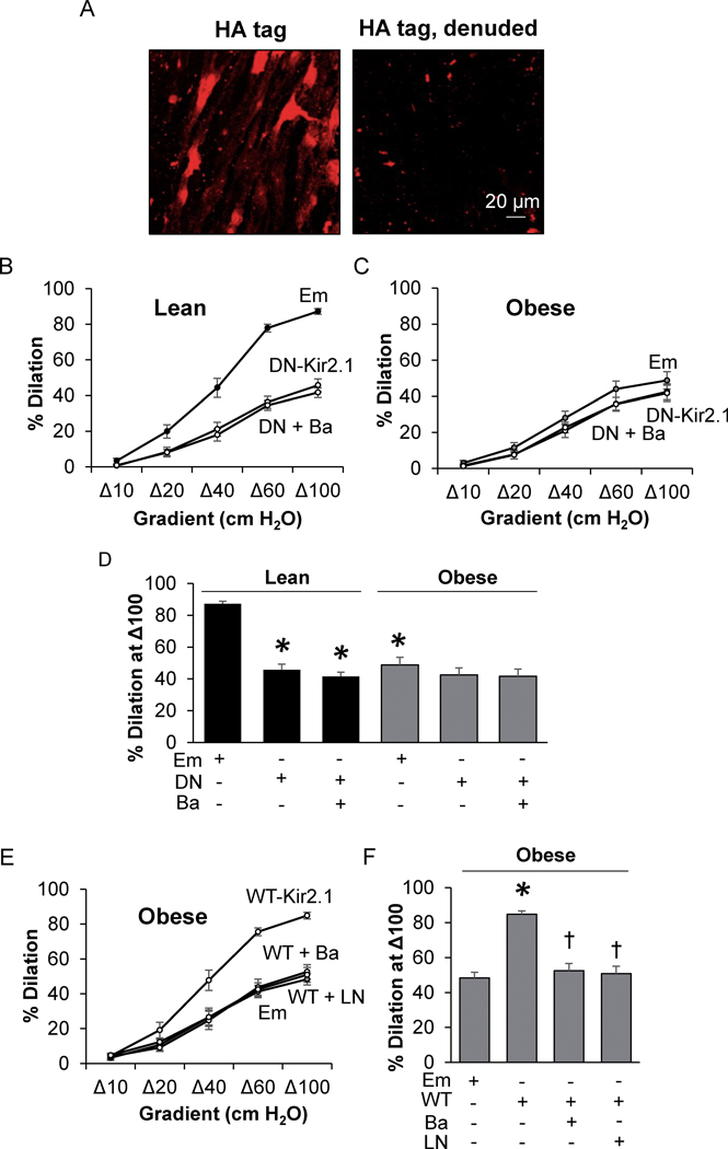
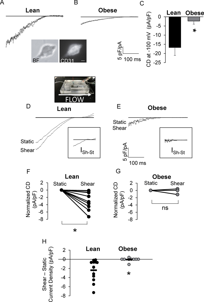
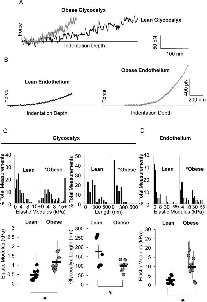
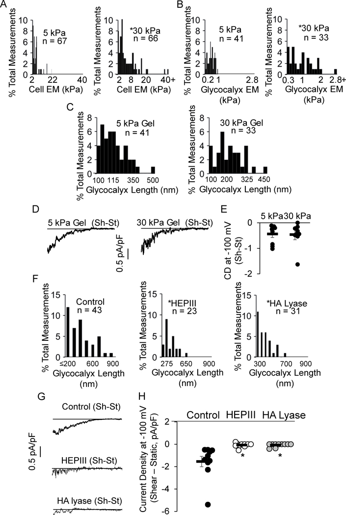
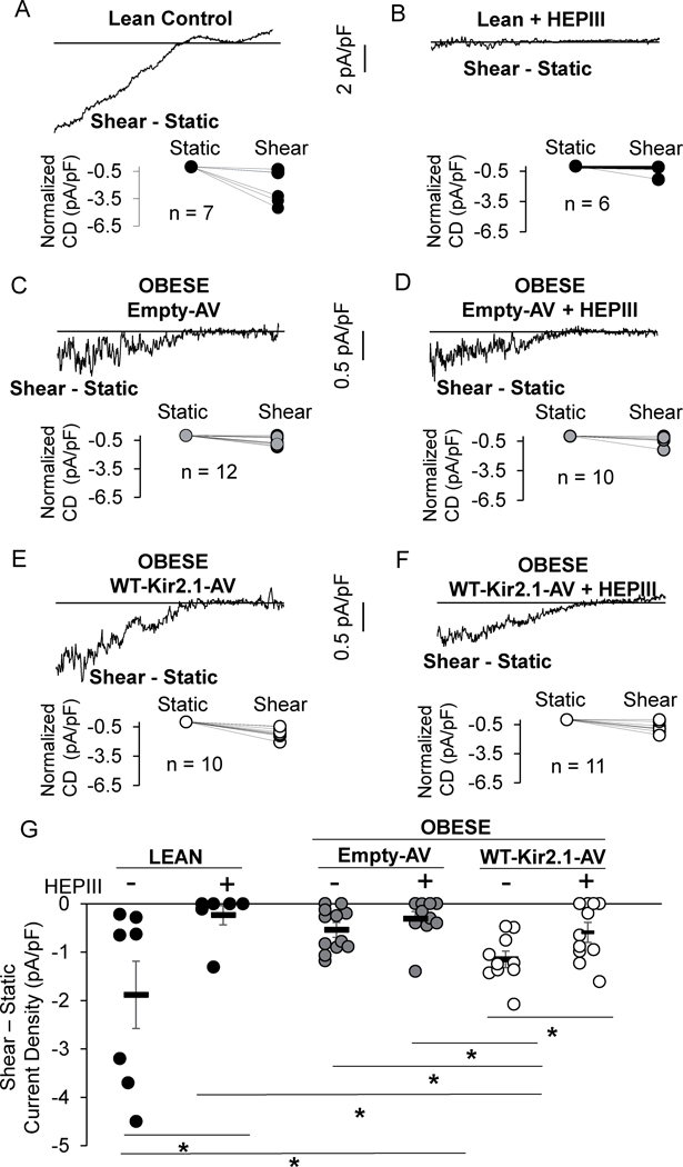
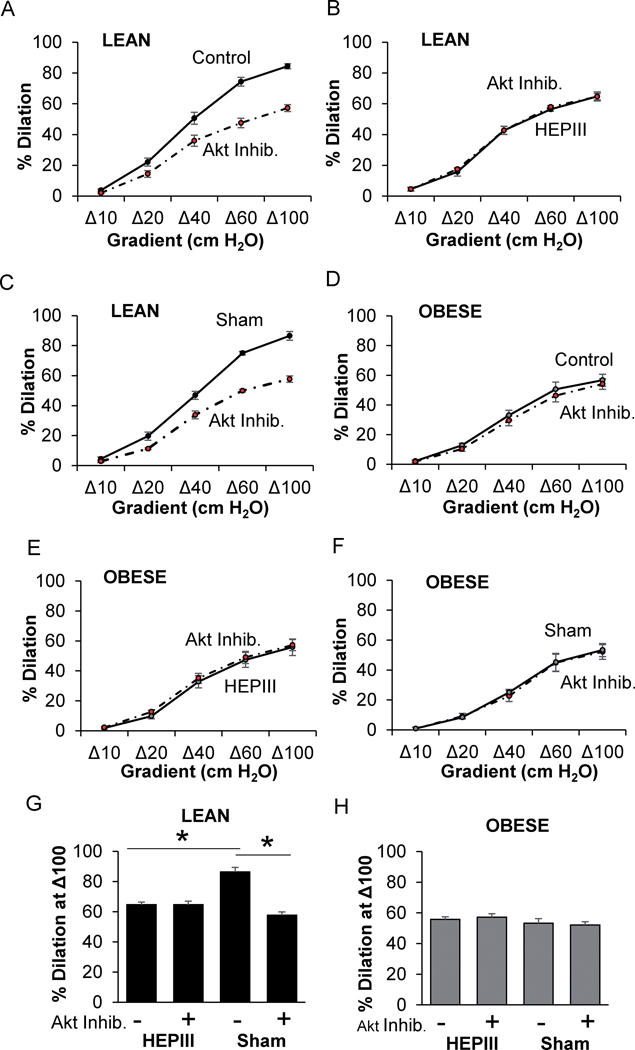
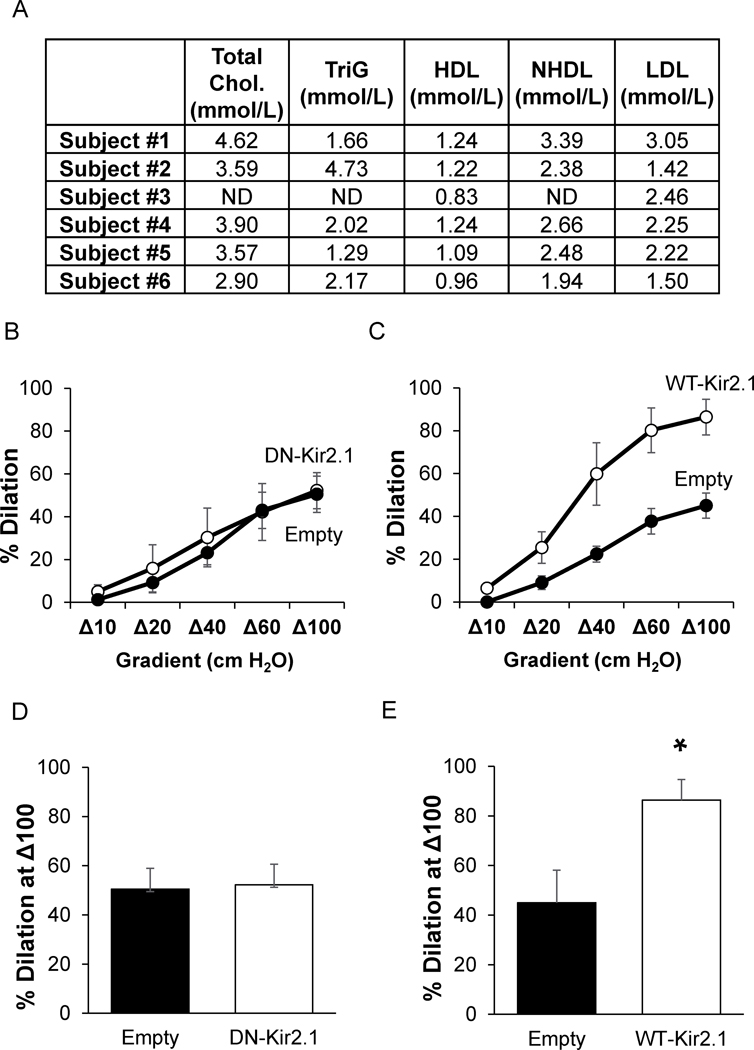
Objective: To determine if endothelial dysfunction in a mouse model of diet-induced obesity and in obese humans is mediated by the suppression of endothelial Kir (inwardly rectifying K+) channels. Approach and Results: Endothelial dysfunction, observed as reduced dilations to flow, occurred after feeding mice a high-fat, Western diet for 8 weeks. The functional downregulation of endothelial Kir2.1 using dominant-negative Kir2.1 construct resulted in substantial reductions in the response to flow in mesenteric arteries of lean mice, whereas no effect was observed in arteries of obese mice. Overexpressing wild-type-Kir2.1 in endothelium of arteries from obese mice resulted in full recovery of the flow response. Exposing freshly isolated endothelial cells to fluid shear during patch-clamp electrophysiology revealed that the flow-sensitivity of Kir was virtually abolished in cells from obese mice. Atomic force microscopy revealed that the endothelial glycocalyx was stiffer and the thickness of the glycocalyx layer reduced in arteries from obese mice. We also identified that the length of the glycocalyx is critical to the flow-activation of Kir. Overexpressing Kir2.1 in endothelium of arteries from obese mice restored flow- and heparanase-sensitivity, indicating an important role for heparan sulfates in the flow-activation of Kir. Furthermore, the Kir2.1-dependent component of flow-induced vasodilation was lost in the endothelium of resistance arteries of obese humans obtained from biopsies collected during bariatric surgery.

Conclusions: We conclude that obesity-induced impairment of flow-induced vasodilation is attributed to the loss of flow-sensitivity of endothelial Kir channels and propose that the latter is mediated by the biophysical alterations of the glycocalyx.

Keywords: Kir channels; endothelial cells; glycocalyx; nitric oxide; obesity; vasodilation.


Figures

Figure 1.
Figure 1.. The loss of Kir2.1 results in FIV impairment in mesenteric adipose arteries of obese mice.
A) Images of intact and denuded en face mesenteric arteries transduced with the VE-Cadherin-WT-Kir2.1-HA tag (WT-Kir2.1)-adenovirus (AV) and stained for the HA-tag. The magnification is 25x. Ex vivo flow-induced vasodilation (FIV) measurements in arteries from B) lean and C) obese mice incubated with Empty (Em)-AV or VE-Cadherin-dominant negative-Kir2.1-AV (DN-Kir2.1) +/− BaCl2 (Ba) (n = 8). D) Analysis of FIV at a flow of Δ100; * indicates significance when compared to lean Em-AV (p<0.05). E) Ex vivo FIV measurements in arteries isolated from obese mice after incubation with either Em-AV or WT-Kir2.1-AV +/− BaCl2 or LNAME (LN) (n = 8). F) Analysis of the %dilation at Δ100; † and * indicate significance when compared to VE-Cadherin-WT-Kir2.1-HA-AV or Em-AV, respectively. Data from an equal number of male and female mice were combined in B-F.
Figure 2.
Figure 2.. Kir channels are impaired in endothelial cells of obese mice.
Representative patch clamp recordings of IKir in endothelial cells (ECs) from A) lean and B) obese mouse mesenteric arteries. Inset: bright field (BF) and CD31 fluorescence image of a freshly isolated EC. Scale bar=5 μm. C) Group data of Kir current density (CD) at −100 mV (*p <0.05). Representative recordings of IKir in ECs from D) lean and E) obese mice before (Static) and after flow (Shear). D: A picture of the parallel plate flow chamber. Insets show the shear-induced increase in IKir for respective recordings. Shear-induced CD for each cell tested from F) lean and G) obese mice normalized to static CD (n = 12 cells from 6 lean mice and 9 cells from 4 obese mice, *p<0.05). H) Group data comparing shear-induced increases in EC IKir (*p<0.05).
Figure 3.
Figure 3.. Obesity results in increased stiffness and decreased thickness of endothelial glycocalyx.
Representative atomic force microscopy force-distance curves generated from A) the glycocalyx and B) endothelium of en face arteries. Group data comparing the C) elastic moduli of the endothelial glycocalyx (left column), thickness of the glycocalyx (right column), and D) elastic moduli of the endothelial layer in arteries from lean and obese mice. For elastic modulus data sets, each data point represents the measurements taken from an individual artery (n = 8 or 9 arteries from 5 mice/group, *p<0.05).
Figure 4.
Figure 4.. Flow-sensitivity of Kir channels critically depends on glycocalyx length.
Histograms generated from A, B) endothelial cell and glycocalyx elastic moduli, and C) glycocalyx length of Human Adipose Microvascular Endothelial Cells (HAMECs) seeded on 5 vs. 30 kPa polyacrylamide gels (*p<0.05). D) Representative recordings and E) group data show flow-induced increase in IKir in HAMECs seeded on gels. F) Histograms detailing the length of the glycocalyx following enzymatic removal of heparan sulfates (middle) and hyaluronic acids (right) (*p<0.05). G) Representative recordings and H) group data show flow-induced increase in IKir following treatment with enzymes (*p<0.05). Data represents 4–5 independent experiments. Shear-Static, Sh-St. heparanase III, HEPIII. Hyaluronic acid lyase, HA lyase.
Figure 5.
Figure 5.. Overexpressing Kir2.1 in endothelium restores Kir flow- and HEPIII-sensitivity.
Representative shear-induced IKir recordings from A) control and B) heparanse III treated (HEPIII) lean mouse endothelial cells (ECs). Representative IKir recordings from ECs isolated from obese mouse mesenteric arteries exposed to C,D) Empty-adenovirus (Em-AV) or E,F) VE-Cadherin-WT-Kir2.1-AV with and without HEPIII treatment. G) Group data (n = 7–12 cells from 4–8 mice/group, *p<0.05). Data from males and female obese mice were combined. Current Density, CD.
Figure 6.
Figure 6.. Flow-mediated HS-glycocalyx-Akt signaling is impaired in obesity.
The effects of Akt inhibition and heparanse III (HEPIII) on flow-induced vasodilation (FIV) in arteries from A-C,G) lean and D-F,H) obese mice are shown (*p<0.05). Sham arteries received a luminal perfusion of buffer without HEPIII (n=4–10 vessels/treatment from 4–5 mice/group). Akt Inhibitor, Akt Inhib. Both male and female mice were included.
Figure 7.
Figure 7.. Kir2.1 does not contribute to FIV in mesenteric adipose arteries of obese humans.
A) The table shows individual subject blood lipids measured by the clinic at the UIC for total cholesterol (Chol.), triglycerides (TriG), high density lipoproteins (HDL), non-HDL (NHDL), and low density lipoproteins (LDL). Flow-induced vasodilation measurements in human arteries exposed to B) VE-Cadherin-dominant negative-Kir2.1-AV (DN-Kir2.1) or C) VE-Cadherin-WT-Kir2.1-HA-AV (WT-Kir2.1). Analysis of the %dilation at a flow of Δ100 for D) DN-Kir2.1 and E) WT-Kir2.1 (*p<0.05). ND – not determined.

References

    1. Stapleton PA, James ME, Goodwill AG and Frisbee JC. Obesity and vascular dysfunction. Pathophysiology. 2008;15:79–89. - PMC - PubMed
    1. Grizelj I, Cavka A, Bian JT, Szczurek M, Robinson A, Shinde S, Nguyen V, Braunschweig C, Wang E, Drenjancevic I and Phillips SA. Reduced flow-and acetylcholine-induced dilations in visceral compared to subcutaneous adipose arterioles in human morbid obesity. Microcirculation. 2015;22:44–53. - PMC - PubMed
    1. Toda N and Okamura T. Obesity impairs vasodilatation and blood flow increase mediated by endothelial nitric oxide: an overview. J Clin Pharmacol. 2013;53:1228–39. - PubMed
    1. Williams IL, Wheatcroft SB, Shah AM and Kearney MT. Obesity, atherosclerosis and the vascular endothelium: mechanisms of reduced nitric oxide bioavailability in obese humans. Int J Obes Relat Metab Disord. 2002;26:754–64. - PubMed
    1. Sharma JN, Al-Omran A and Parvathy SS. Role of nitric oxide in inflammatory diseases. Inflammopharmacology. 2007;15:252–9. - PubMed

Publication types

MeSH terms