. 2020 Jul 22;11(1):306.
doi: 10.1186/s13287-020-01814-4.

METTL1-mediated m7G methylation maintains pluripotency in human stem cells and limits mesoderm differentiation and vascular development

Affiliations

METTL1-mediated m7G methylation maintains pluripotency in human stem cells and limits mesoderm differentiation and vascular development

Yujie Deng et al. Stem Cell Res Ther. .

Abstract

Background: 7-Methylguanosine (m7G) is one of the most conserved modifications in nucleosides within tRNAs and rRNAs. It plays essential roles in the regulation of mRNA export, splicing, and translation. Recent studies highlighted the importance of METTL1-mediated m7G tRNA methylome in the self-renewal of mouse embryonic stem cells (mESCs) through its ability to regulate mRNA translation. However, the exact mechanisms by which METTL1 regulates pluripotency and differentiation in human induced pluripotent stem cells (hiPSCs) remain unknown. In this study, we evaluated the functions and underlying molecular mechanisms of METTL1 in regulating hiPSC self-renewal and differentiation in vivo and in vitro.

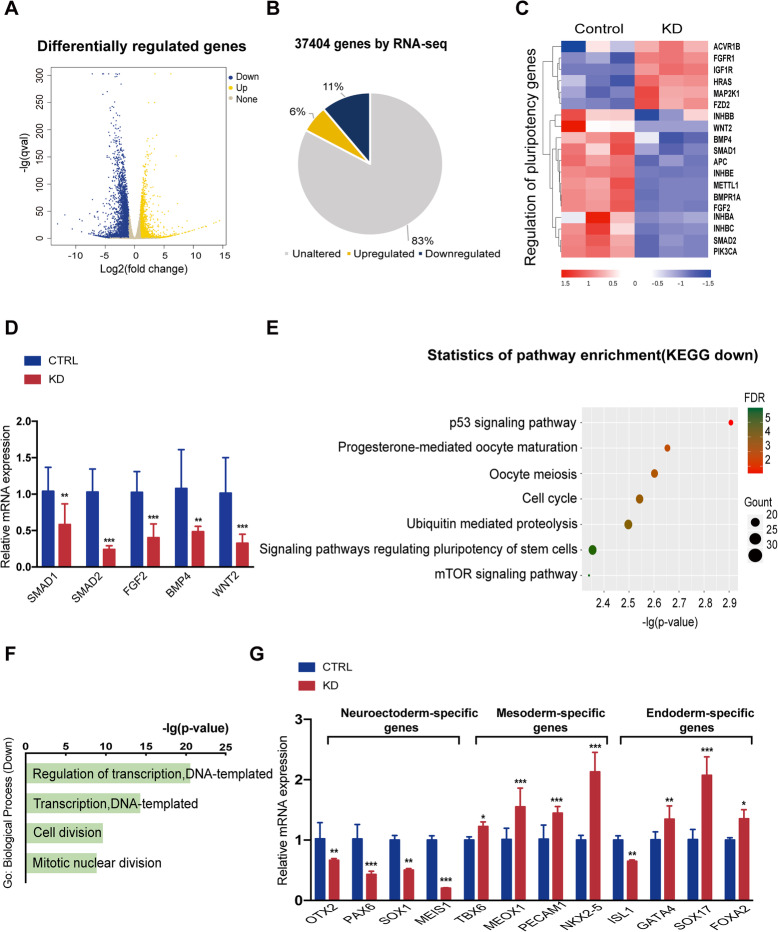
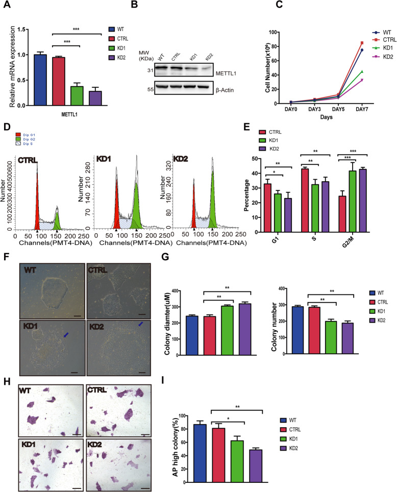
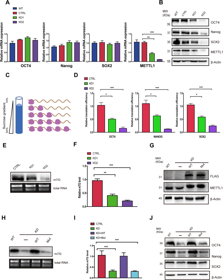
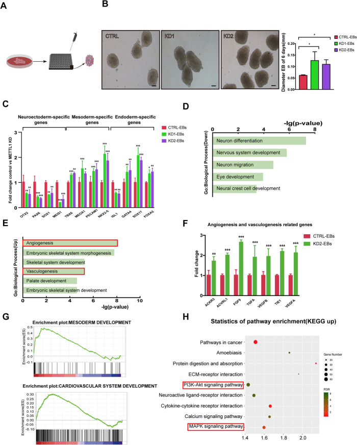
Methods: By establishing METTL1 knockdown (KD) hiPSCs, gene expression profiling was performed by RNA sequencing followed by pathway analyses. Anti-m7G northwestern assay was used to identify m7G modifications in tRNAs and mRNAs. Polysome profiling was used to assess the translation efficiency of the major pluripotent transcription factors. Moreover, the in vitro and in vivo differentiation capacities of METTL1-KD hiPSCs were assessed in embryoid body (EB) formation and teratoma formation assays.

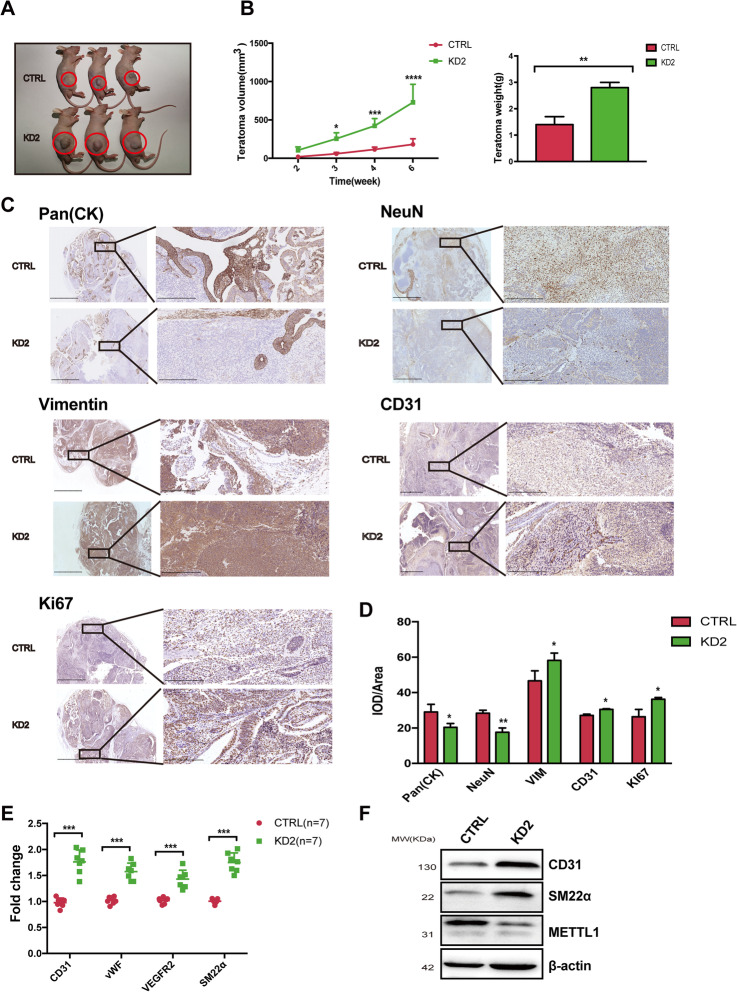
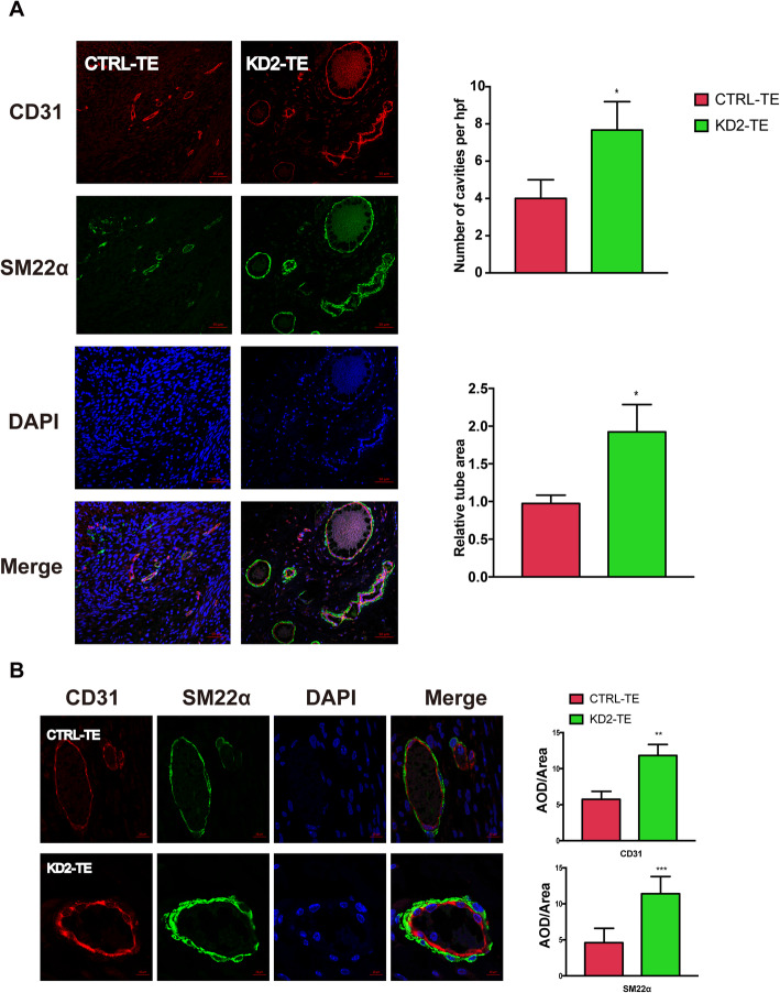
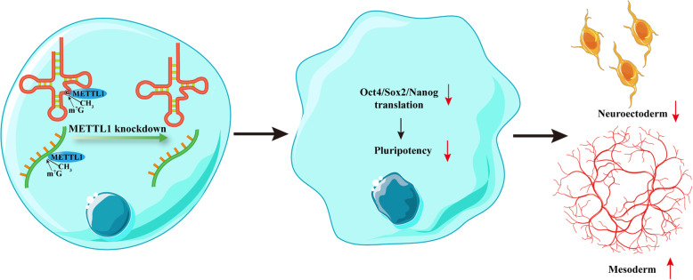
Results: METTL1 silencing resulted in alterations in the global m7G profile in hiPSCs and reduced the translational efficiency of stem cell marker genes. METTL1-KD hiPSCs exhibited reduced pluripotency with slower cell cycling. Moreover, METTL1 silencing accelerates hiPSC differentiation into EBs and promotes the expression of mesoderm-related genes. Similarly, METTL1 knockdown enhances teratoma formation and mesoderm differentiation in vivo by promoting cell proliferation and angiogenesis in nude mice.

Conclusion: Our findings provided novel insight into the critical role of METTL1-mediated m7G modification in the regulation of hiPSC pluripotency and differentiation, as well as its potential roles in vascular development and the treatment of vascular diseases.

Keywords: Differentiation; Human induced pluripotent stem cells (hiPSCs); Mesoderm; N7-methylguanosine (m7G); Pluripotency; Vasculogenesis.


Conflict of interest statement

The authors declare that they have no competing interests

Figures

Fig. 1
Fig. 1
Identification of METTL1-regulated genes by RNA-seq analysis in hiPSCs. HiPSCs were transduced with shRNAs-METTL1, and pooled clones were selected. a Volcano plot illustrating differentially expressed genes (DEGs) between control and METTL1 knockdown (KD) hiPSCs. Genes upregulated and downregulated are shown in yellow and blue, respectively. Values are presented as the log2 of tag counts. b A total of 37,404 genes were differentially expressed, of which 2301 (6%) were upregulated and 4125 (11%) were downregulated in METTL1-KD hiPSCs. c Heatmap showing DEGs involved in stem cells pluripotency pathways. Each lane corresponds to an independent biological sample. Scale bar: log2 FPKM. d qRT-PCR analysis assessing the mRNA levels of genes involved in pluripotency, in control and METTL1-KD hiPSCs. e KEGG pathway analysis of genes downregulated in METTL1-KD hiPSCs. f Gene ontology (GO) functional clustering of genes downregulated in METTL1-KD hiPSCs; the top 4 most significant biological processes are shown. g qRT-PCR validation analysis shows the mRNA expression fold change of neuroectoderm-, endoderm- and mesoderm-specific genes in METTL1-KD vs. control hiPSCs. Data are presented as the mean ± SEM from three independent experiments. *P < 0.05; **P < 0.01; ***P < 0.001
Fig. 2
Fig. 2
METTL1 is essential for hiPSC self-renewal and pluripotency. Different METTL1-KD hiPSC single-cell clones were selected, and two clones were used for further analyses. a qRT-PCR analysis of METTL1 mRNA expression in control and METTL1-KD hiPSC. b Western blot of the control and METTL1-KD samples with indicated antibodies. c Cell proliferation in control and METTL1-KD hiPSC. The cell numbers were quantified after 3, 5, and 7 days of culture; n = 3. d, e Cell cycle analysis in METTL1-KD and control hiPSC. f, g Colony formation assay of control and METTL1-KD hiPSC. Colony size and numbers were measured at day 7. f Representative images of the colonies. Scale bar, 100 μm. g Colony diameter and number quantification. h, i Alkaline phosphatase (AP) staining of control and METTL1-KD colonies. h Representative images; scale bar, 100 μm. i Percentage of AP positive colonies. The data are presented as mean ± SEM; n = 3; *P < 0.05; **P < 0.01; ***P < 0.001
Fig. 3
Fig. 3
METTL1-mediated m7G methylation is required for OCT4, Nanog, and SOX2 translation in hiPSCs. a, b Effects of METTL1 KD on stem cell marker expression. a qRT-PCR analysis assessing the mRNA levels of OCT4, Nanog, and SOX2 in control and METTL1-KD hiPSCs. b Western blot showing OCT4, Nanog, and SOX2 protein levels in control and METTL1-KD hiPSCs. c, d Effects of METTL1-KD on translational efficiency of stem cell markers. c Schematic representation of the sucrose gradient procedure followed to isolate ribosome-free and ribosome-bound RNAs. d Total and polysome-fractionated RNAs from control and METTL1-KD cells were quantified by qRT-PCR, and translational efficiency was presented as relative percent of polysome associated mRNA to input mRNA of indicated stem cell markers. e Anti-m7G Northwestern blots (upper panel). RNAs were separated on TBE-urea gels, transferred to nylon membranes, and probed with anti-m7G antibodies. Expression was compared to total RNA controls (lower panel). f Quantification of relative m7G levels. g, h Rescue of stem cell markers expression by the exogenous expression of METTL1-WT but not an enzyme-inactive mutant (Mut). g Western blot analysis of reconstituted hiPSCs. h Anti-m7G Northwestern blot of m7G modifications (upper panel) and agarose gel electrophoresis of total RNA (lower panel) in METTL1-mutant samples. i Quantification of m7G levels. j Rescue of stem cell marker expression assessed by WB analysis. Data are presented as mean ± SEM; n = 3; **P < 0.01; ***P < 0.001
Fig. 4
Fig. 4
METTL1 silencing accelerates embryonic body formation and increases the expression of angiogenesis-related genes. a Schematic diagram showing the EB spheroid formation process. Cells were seeded on ultralow attachment 96-well spheroid microplates to induce EB formation. b Representative images and quantification of EB formation in control and METTL1-KD hiPSCs on day 6. Scale bar, 50 μm. c qRT-PCR analysis assessing the mRNA levels in control and METTL1-KD EBs. d, e Gene ontology analysis showing functional enrichment in biological process in genes downregulated and upregulated in METTL1-KD EBs. f qRT-PCR analysis assessing the mRNA levels of angiogenesis- and vasculogenesis-related genes in control and METTL1-KD EBs. g GSEA showing enrichment of the mesoderm and cardiovascular development gene signatures. Y-axis indicates enrichment score (ES), while the X-axis indicates positively and negatively correlated gene sets. h KEGG pathway analysis of genes upregulated in METTL1-KD EBs. Data are presented as mean ± SEM; n = 3; *P < 0.05, **P < 0.01; ***P < 0.001
Fig. 5
Fig. 5
METTL1 knockdown accelerates teratoma development and angiogenesis in nude mice. a Teratomas formed 6 weeks post-injection in BALB/C nude mice. b Teratoma weight and volume were quantified; n = 5. c, d Immunohistochemical analysis of teratomas from METTL1-KD or control hiPSCs. Scale bar, 2.5 mm and 250 μm, respectively. Quantification of cell number is shown. e Quantification of CD31, vWF, VEGFR2, and SM22α mRNA levels in teratomas from METTL1-KD and control hiPSCs; n = 7 for tumor and control samples. f Representative western blot images for CD31, METTL1, and SM22α protein levels in teratomas from METTL1-KD hiPSCs and control hiPSCs; n = 3. Data are presented as mean ± SEM; *P < 0.05; ***P < 0.001; ****P < 0.0001
Fig. 6
Fig. 6
METTL1 silencing promotes angiogenesis and increased CD31 and SM22α expression in teratomas. a Representative images from control and METTL1-KD2 teratomas stained for CD31 (red), and SM22α (green); nuclei were counterstained with DAPI (blue). The number of cavities per hpf and relative tube area were determined using ImageJ software. Scale bar, 50 μm. b Representative images of vessels at high magnification (× 100) and quantification of CD31 and SM22α intensities in control and METTL1-KD teratomas; nuclei were counterstained with DAPI. Scale bar, 10 μm. Data are presented as mean ± SEM; n = 3; **P < 0.01, ***P < 0.001
Fig. 7
Fig. 7
A graphic for the role of METTL1 in regulating stem cell pluripotency and differentiation. METTL1-mediated m7G methylation is essential for the translation of pluripotency transcription factors. METTL1 knockdown impairs neurectoderm formation while accelerates mesoderm differentiation and vasculogenesis/angiogenesis

References

    1. Takahashi K, Tanabe K, Ohnuki M, Narita M, Ichisaka T, Tomoda K, Yamanaka S. Induction of pluripotent stem cells from adult human fibroblasts by defined factors. Cell. 2007;131(5):861–872. - PubMed
    1. Yu J, Vodyanik MA, Smuga-Otto K, Antosiewicz-Bourget J, Frane JL, Tian S, Nie J, Jonsdottir GA, Ruotti V, Stewart R, et al. Induced pluripotent stem cell lines derived from human somatic cells. Science. 2007;318(5858):1917–1920. - PubMed
    1. Wu SM, Hochedlinger K. Harnessing the potential of induced pluripotent stem cells for regenerative medicine. Nat Cell Biol. 2011;13(5):497–505. - PMC - PubMed
    1. Stadtfeld M, Hochedlinger K. Induced pluripotency: history, mechanisms, and applications. Genes Dev. 2010;24(20):2239–2263. - PMC - PubMed
    1. Thomson M, Liu SJ, Zou LN, Smith Z, Meissner A, Ramanathan S. Pluripotency factors in embryonic stem cells regulate differentiation into germ layers. Cell. 2011;145(6):875–889. - PMC - PubMed

Publication types

LinkOut - more resources