. 2021 Feb;42(2):301-310.
doi: 10.1038/s41401-020-0478-3. Epub 2020 Jul 22.

Artesunate synergizes with sorafenib to induce ferroptosis in hepatocellular carcinoma

Affiliations

Artesunate synergizes with sorafenib to induce ferroptosis in hepatocellular carcinoma

Zhong-Jie Li et al. Acta Pharmacol Sin. 2021 Feb.

Abstract

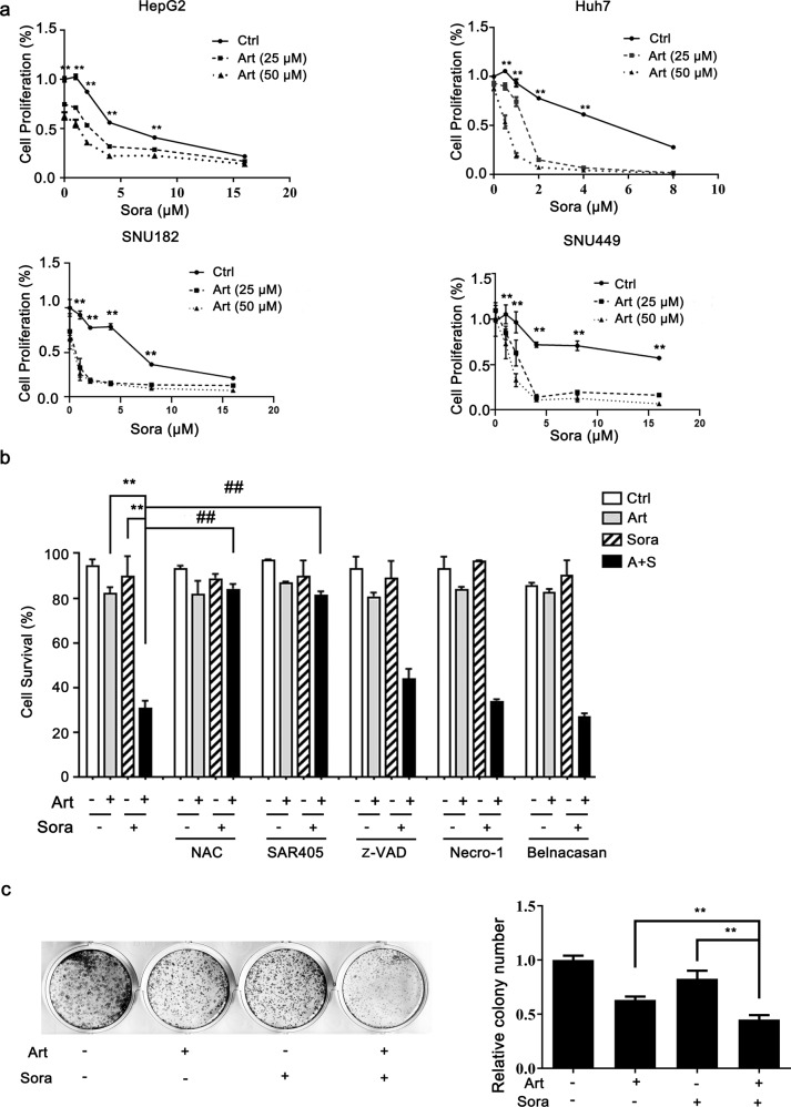
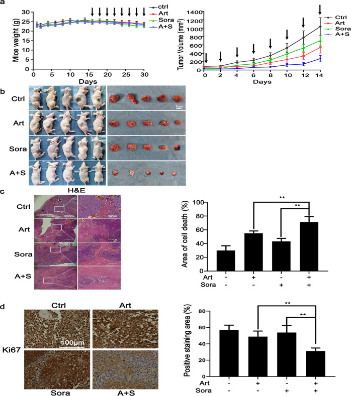
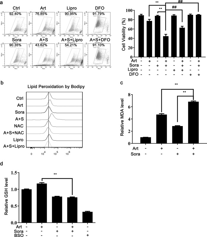
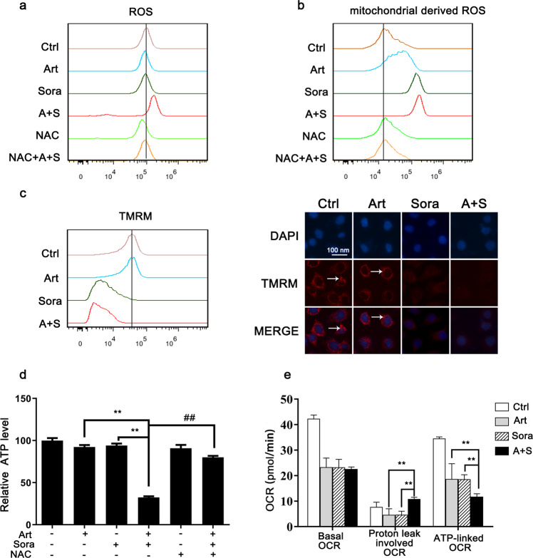
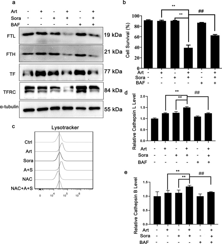
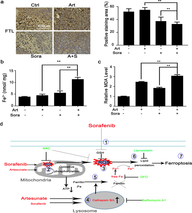
Sorafenib is the first-line medication for advanced hepatocellular carcinoma (HCC), but it can only extend limited survival. It is imperative to find a combination strategy to increase sorafenib efficacy. Artesunate is such a preferred candidate, because artesunate is clinically well-tolerated and more importantly both drugs can induce ferroptosis through different mechanisms. In this study we investigated the combined effect of sorafenib and artesunate in inducing ferroptosis of HCC and elucidated the involved molecular mechanisms. We showed that artesunate greatly enhanced the anticancer effects of low dose of sorafenib against Huh7, SNU-449, and SNU-182 HCC cell lines in vitro and against Huh7 cell xenograft model in Balb/c nude mice. The combination index method confirmed that the combined effect of sorafenib and artesunate was synergistic. Compared with the treatment with artesunate or sorafenib alone, combined treatment induced significantly exacerbated lipid peroxidation and ferroptosis, which was blocked by N-acetyl cysteine and ferroptosis inhibitors liproxstatin-1 and deferoxamine mesylate, but not by inhibitors of other types of cell death (z-VAD, necrostatin-1 and belnacasan). In Huh7 cells, we demonstrated that the combined treatment induced oxidative stress and lysosome-mediated ferritinophagy, two essential aspects of ferroptosis. Sorafenib at low dose mainly caused oxidative stress through mitochondrial impairments and SLC7A11-invovled glutathione depletion. Artesunate-induced lysosome activation synergized with sorafenib-mediated pro-oxidative effects by promoting sequential reactions including lysosomal cathepsin B/L activation, ferritin degradation, lipid peroxidation, and consequent ferroptosis. Taken together, artesunate could be repurposed to sensitize sorafenib in HCC treatment. The combined treatment can be easily translated into clinical applications.

Keywords: Artesunate; Ferroptosis; HCC; Lysosome; Mitochondria; Sorafenib.


Conflict of interest statement

The authors declare no competing interests.

Figures

Fig. 1
Fig. 1. Artesunate sensitized HCC cells to sorafenib-induced death.
a HepG2, Huh7 and SNU-449 cells were treated with artesunate (Art) and/or sorafenib (Sora) as indicated for 24 h, while SNU-182 cells were administered the same treatments for 12 h. Cell proliferation was measured by the MTT assay. b Huh7 cells were treated with Art (25 μM) and/or Sora (2 μM) in the presence or absence of pretreatment with different inhibitors for 24 h. These inhibitors included NAC (an ROS scavenger, 5 mM), SAR405 (an inhibitor of PIK3C3/Vps34 and consequent autophagy, 1 µM), z-VAD (a pan-caspase inhibitor, 40 μM), necrostatin-1 (an inhibitor of RIP kinase and consequent necroptosis, 20 µM), and belnacasan (an inhibitor of caspase-1 and consequent pyroptosis, 20 µM). The cell survival rate was determined by the PI exclusion assay. c Huh7 cells were seeded in 12-well plates and treated with Art (10 μM) and Sora (1 μM) for 10 days. A + S, the combination of Art and Sora; **P < 0.01 compared with Art alone or Sora alone; ##P < 0. 01 compared with the combination of Art and Sora.
Fig. 2
Fig. 2. The combined treatment inhibited xenograft tumors in vivo through extensive cell death.
a Once the size of the inoculated xenograft nodules reached 80–100 mm3, the mice were randomly divided into four groups (n = 5 per group) and treated by gavage with Art (30 mg/kg body weight), Sora (20 mg/kg), the combination of Art and Sora or PBS (control) every other day. The weight of the mice (left panel) and tumor growth (right panel) were monitored regularly. The arrows indicate drug treatment. b The mice were sacrificed, and the tumors were resected for subsequent experiments. c Xenograft tumor sections were stained with H&E. The area of cell death was indicated by smeared cell morphology and faint nuclear staining. The area of cell death is presented in the right panel. d Tumor cell proliferation status was analyzed by IHC staining for Ki-67. **P < 0.01.
Fig. 3
Fig. 3. The combined treatment-induced lipid peroxidation and ferroptosis.
a Huh7 cells were treated as indicated with or without Liproxstatin-1 (Lipro, 10 μM) and deferoxamine (DFO, 100 μM). The cell survival rate was quantified by the PI exclusion assay. b Huh7 cells were treated as indicated with or without Lipro or NAC. The level of lipid peroxidation was determined with BODIPY 581/591 C11. After cell treatments, MDA production (c) and GSH content (d) were measured with respective assay kits as described. BSO (10 μM) was used as a positive control. **P < 0.01.
Fig. 4
Fig. 4. The combined treatment impaired mitochondrial functions.
Huh7 cells were treated as indicated in Fig. 3b. Then, the levels of ROS (a) and mitochondrial-derived ROS (b) were measured with DCFDA and MitoSOX Red, respectively. c The mitochondrial membrane potential was determined with TMRM by using flow cytometry (left panel) and fluorescence microscopy (right panel). d The cellular ATP levels were determined by biochemical assays after the indicated treatments. e Huh7 cells were washed and recultured in fresh XF medium with the indicated drugs for 1 h. Then, the mitochondrial respiratory OCR was determined by Seahorse metabolic assay. Basal respiration OCR, proton leak-involved OCR and ATP-linked OCR are summarized. OCR oxygen consumption rate. **P < 0.01.
Fig. 5
Fig. 5. The combined treatment-induced lysosomal activation and ferritin degradation.
a Huh7 cells were treated as indicated with or without bafilomycin A1 (100 nM) for 12 h before Western blotting. b Huh7 cells were treated as indicated in (a) for 24 h before the cell survival rate was quantified. c Huh7 cells were treated as indicated in Fig. 4a before lysosomal activity analysis using LysoTracker Red. d, e Huh7 cells were treated as indicated with or without bafilomycin A1 (100 nM) pretreatment for 12 h. Then, the lysosomal enzyme activities of cathepsin L (d) and cathepsin B (e) were quantified. **P < 0.01. ##P < 0.01.
Fig. 6
Fig. 6. The combined treatments caused consistent changes in FTL degradation and MDA production in xenograft nodules in vivo.
a Xenograft nodules were collected as shown in Fig. 2b. FTL protein expression was determined by IHC staining as shown in Fig. 2d. The contents of free Fe2+ ions (b) and MDA (c) in tumor nodules were quantified with respective biochemical assay kits. **P < 0.01. d The cartoon describes the molecular mechanisms through which the combined treatment led to ferroptosis. Sorafenib directly inhibits ① GSH synthesis and ② mitochondrial functions, which contributes to ③ extensive oxidative stress. Artesunate primarily ④ promotes lysosomal activation and then synergizes with sorafenib to activate cathepsin B/L activities followed by ⑤ ferritin degradation. The convergence of sorafenib-induced oxidative stress and artesunate-mediated ferritinophagy causes ⑥ lipid peroxidation and eventually ⑦ ferroptosis. DFO, deferoxamine mesylate; GSH glutathione, NAC N-acetyl cysteine; ROS reactive oxygen species. The red color indicates ferroptosis inducers or essential aspects of ferroptosis. The green color indicates inhibitors of ferroptosis induction.

References

    1. Villanueva A, Hernandez-Gea V, Llovet JM. Medical therapies for hepatocellular carcinoma: a critical view of the evidence. Nat Rev Gastroenterol Hepatol. 2012;10:34–42. doi: 10.1038/nrgastro.2012.199. - DOI - PubMed
    1. European Association for the Study of the Liver. EASL clinical practice guidelines: management of hepatocellular carcinoma. J Hepatol. 2018;69:182–236. doi: 10.1016/j.jhep.2018.03.019. - DOI - PubMed
    1. Llovet JM, Ricci S, Mazzaferro V, Hilgard P, Gane E, Blanc JF, et al. Sorafenib in advanced hepatocellular carcinoma. N Engl J Med. 2008;359:378–90. doi: 10.1056/NEJMoa0708857. - DOI - PubMed
    1. Cheng AL, Kang YK, Chen Z, Tsao CJ, Qin S, Kim JS, et al. Efficacy and safety of sorafenib in patients in the Asia-Pacific region with advanced hepatocellular carcinoma: a phase III randomised, double-blind, placebo-controlled trial. Lancet Oncol. 2009;10:25–34. doi: 10.1016/S1470-2045(08)70285-7. - DOI - PubMed
    1. Llovet JM, Montal R, Sia D, Finn RS. Molecular therapies and precision medicine for hepatocellular carcinoma. Nat Rev Clin Oncol. 2018;15:599–616. doi: 10.1038/s41571-018-0073-4. - DOI - PubMed

MeSH terms