

Uveal Melanoma: A Review of the Literature
- PMID: 32700136
- PMCID: PMC7359963
- DOI: 10.1007/s40487-018-0056-8
Uveal Melanoma: A Review of the Literature
Erratum in
-
Correction to: Uveal Melanoma: A Review of the Literature.Oncol Ther. 2019 Jun;7(1):93. doi: 10.1007/s40487-019-0093-y. Oncol Ther. 2019. PMID: 32700195 Free PMC article.
Abstract
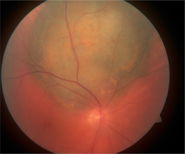
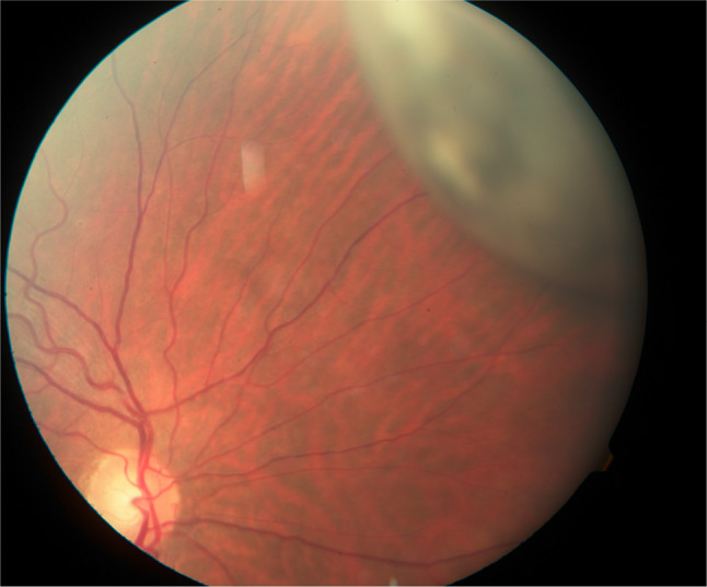
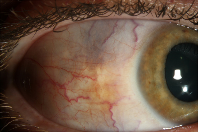
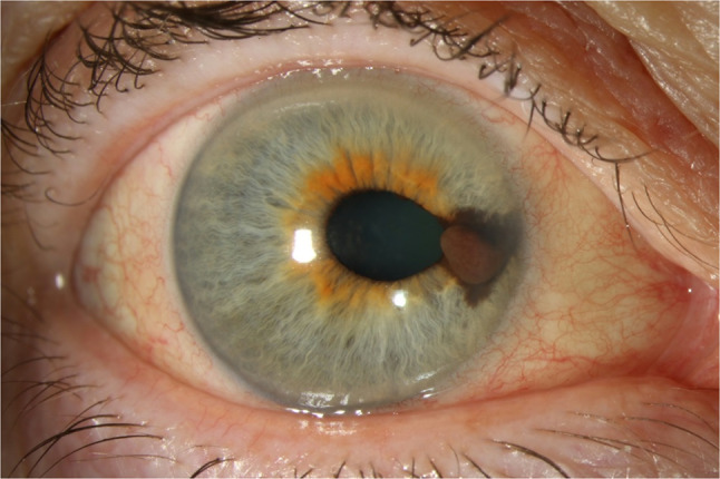
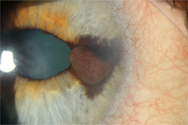
Melanomas affecting different components of the uvea occur with differing frequencies and clinical presentations. Uveal melanoma is diagnosed via funduscopic exam and ancillary tests. These lesions may present with visual findings or incidental findings on physical exam. Metastasis occurs in approximately half of all patients with primary uveal melanoma. The liver is the most common site of metastasis. Enucleation was at one time considered the definitive local treatment for primary uveal melanoma, but has been largely replaced by other therapeutic procedures that aim to prevent metastasis while preserving vision. Unfortunately, metastasis of uveal melanoma almost always proves to be fatal. The current treatment of metastatic uveal melanoma is limited by the intrinsic resistance of uveal melanoma to conventional systemic therapies. Advancements in molecular biology have resulted in the identification of a number of promising prognostic and therapeutic targets. Early detection and therapy are important factors in disease survival. It is imperative that the treating physician be familiar with the clinical features of uveal melanoma and distinguish it from mimickers in order to ensure effective and timely treatment.
Keywords: Choroid; Ciliary body; Iris; Melanoma; Review.
Figures
References
-
- Spagnolo F, Caltabiano G, Queirolo P. Uveal melanoma. Cancer Treat Rev. 2012;38:549–553. - PubMed
-
- Yonekawa Y, Kim IK. Epidemiology and management of uveal melanoma. Hematol Oncol Clin N Am. 2012;26:1170–1184. - PubMed
-
- Nathan P, Cohen V, Coupland S, et al. Uveal melanoma UK national guidelines. Eur J Cancer. 2015. 10.1016/j.ejca.2015.07.013. - PubMed
-
- Shields CL, Manalac J, Das C, Ferguson K, Shields JA. Choroidal melanoma: clinical features, classification, and top 10 pseudomelanomas. Curr Opin Ophthalmol. 2014;25:177–185. - PubMed
LinkOut - more resources
Full Text Sources
Other Literature Sources
Medical
