. 2021 Jun;15(3):1469-1482.
doi: 10.1007/s11682-020-00346-y.

Early deafness leads to re-shaping of functional connectivity beyond the auditory cortex

Affiliations

Early deafness leads to re-shaping of functional connectivity beyond the auditory cortex

Kamil Bonna et al. Brain Imaging Behav. 2021 Jun.

Abstract

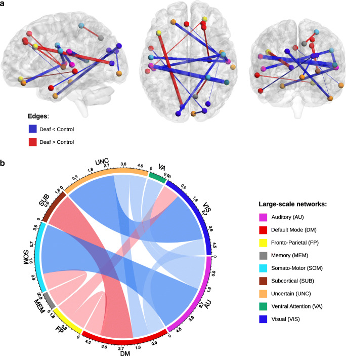
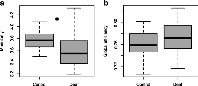
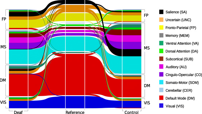
Early sensory deprivation, such as deafness, shapes brain development in multiple ways. Deprived auditory areas become engaged in the processing of stimuli from the remaining modalities and in high-level cognitive tasks. Yet, structural and functional changes were also observed in non-deprived brain areas, which may suggest the whole-brain network changes in deaf individuals. To explore this possibility, we compared the resting-state functional network organization of the brain in early deaf adults and hearing controls and examined global network segregation and integration. Relative to hearing controls, deaf adults exhibited decreased network segregation and an altered modular structure. In the deaf, regions of the salience network were coupled with the fronto-parietal network, while in the hearing controls, they were coupled with other large-scale networks. Deaf adults showed weaker connections between auditory and somatomotor regions, stronger coupling between the fronto-parietal network and several other large-scale networks (visual, memory, cingulo-opercular and somatomotor), and an enlargement of the default mode network. Our findings suggest that brain plasticity in deaf adults is not limited to changes in the auditory cortex but additionally alters the coupling between other large-scale networks and the development of functional brain modules. These widespread functional connectivity changes may provide a mechanism for the superior behavioral performance of the deaf in visual and attentional tasks.

Keywords: Brain plasticity; Deafness; Functional connectivity; Graph theory; Resting-state fMRI.


Conflict of interest statement

The authors declare no competing interests

Figures

Fig. 1
Fig. 1
Edge-wise functional network differences visualized (a) in brain space and (b) as a chord diagram. (a) Connections that are significantly stronger (red) or weaker (blue) in deaf adults. Edge thickness reflects t-test statistic strength. (b) Chord diagram representing the number of significant edges between different large-scale networks. Red bands represent edges with stronger functional connectivity in the deaf compared to hearing control, while blue bands represent edges with weaker functional connectivity
Fig. 2
Fig. 2
Differences in graph measures of cortical segregation and integration between deaf adults and the control group. (a) The difference in network segregation measured as modularity. (b) The difference in network integration measured as global efficiency. Boxplots represent topological values calculated for 25% threshold. * p < 0.05
Fig. 3
Fig. 3
An alluvial diagram representing the segregation of group-averaged networks using a data-driven approach in the deaf (left side of the diagram) and the control group (right side of the diagram). This segregation is then compared against a priori segregation into 13 well-known networks based on meta-analysis studies (Power et al. 2011), shown in the middle column, and described in the right-hand side legend. Note that salience nodes (black) are part of the fronto-parietal (FP) module in the deaf group but fall into the multi-system (MS) module in the control group. Also, the ventral-attention nodes (dark green) are part of the MS module in the control group, but in the deaf group, they are part of the default mode module (DM). The composition of the last visual module (VIS) is highly consistent in both groups

References

    1. Alavash M, Tune S, Obleser J. Modular reconfiguration of an auditory control brain network supports adaptive listening behavior. Proceedings of the National Academy of Sciences of the United States of America. 2019;116(2):660–669. doi: 10.1073/pnas.1815321116. - DOI - PMC - PubMed
    1. Alexander-Bloch AF, Gogtay N, Meunier D, Birn R, Clasen L, Lalonde F, Lenroot R, Giedd J, Bullmore ET. Disrupted modularity and local connectivity of brain functional networks in childhood-onset schizophrenia. Frontiers in Systems Neuroscience. 2010;4:147. doi: 10.3389/fnsys.2010.00147. - DOI - PMC - PubMed
    1. Allen JS, Emmorey K, Bruss J, Damasio H. Morphology of the insula in relation to hearing status and sign language experience. The Journal of Neuroscience: The Official Journal of the Society for Neuroscience. 2008;28(46):11900–11905. doi: 10.1523/JNEUROSCI.3141-08.2008. - DOI - PMC - PubMed
    1. Ashburner J, Friston KJ. Unified segmentation. NeuroImage. 2005;26(3):839–851. doi: 10.1016/j.neuroimage.2005.02.018. - DOI - PubMed
    1. Auer ET, Jr, Bernstein LE, Sungkarat W, Singh M. Vibrotactile activation of the auditory cortices in deaf versus hearing adults. Neuroreport. 2007;18(7):645–648. doi: 10.1097/WNR.0b013e3280d943b9. - DOI - PMC - PubMed