Clinical and Radiographic Gingival Thickness Assessment at Mandibular Incisors: an Ex Vivo Study
- PMID: 32700514
- PMCID: PMC11654594
- DOI: 10.3290/j.ohpd.a44925
Clinical and Radiographic Gingival Thickness Assessment at Mandibular Incisors: an Ex Vivo Study
Abstract
Purpose: Gingival phenotype influences the outcomes of various dental procedures. The objective of the current study was to assess the agreement between various clinical and radiographic methods for evaluating gingival thickness.
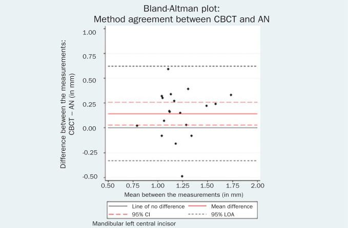
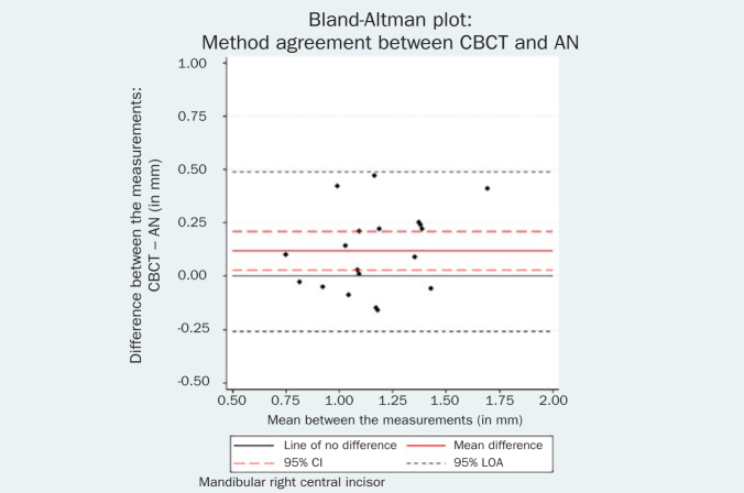
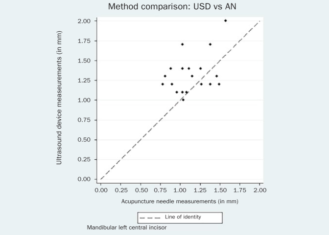
Materials and methods: This ex-vivo study evaluated gingival thickness on 20 porcine cadavers. Gingival thickness was assessed at both central mandibular incisors with: a) trans-gingival probing with a standard periodontal probe (PB); b) trans-gingival probing with a stainless steel acupuncture needle (AN); c) ultrasound device (USD); and d) Cone Beam Computed Tomography (CBCT). Intra-examiner reproducibility and method error were also evaluated.
Results: Trans-gingival measurements with the standard PB and the AN were found to be almost identical in gingival thickness assessment (mean GT 1.11 mm vs 1.14 mm for the left incisor and mean GT 1.12 mm vs 1.11 mm for the right incisor, respectively). USD and CBCT yielded values that were statistically significantly higher than AN. Both USD and CBCT values were higher than PB, but this difference was statistically significant only for the left central incisor. Finally, USD values exceeded CBCT measurements, but this difference was not statistically significant. There was no evidence of systematic differences between the repeated CBCT measurements (p = 0.06 for the left incisor and p = 0.55 for the right incisor).
Conclusions: CBCT measurements proved to be highly repeatable and comparable to the USD measurements, while there were some indications that both CBCT and USD measurements were systematically higher than either PB or AN.
Keywords: cone-beam computed tomography; gingival biotype; periodontal tissue; ultrasound.
Figures


References
-
- Amid R, Mirakhori M, Safi Y, Kadkhodazadeh M, Namdari M. Assessment of gingival biotype and facial hard/soft tissue dimensions in the maxillary anterior teeth region using cone beam computed tomography. Arch Oral Biol. 2017;79:1–6. - PubMed
-
- Anderegg CR, Metzler DG, Nicoll BK. Gingiva thickness in guided tissue regeneration and associated recession at facial furcation defects. J Periodontol. 1995;66:397–402. - PubMed
-
- Baldi C, Pini-Prato G, Pagliaro U, Nieri M, Saletta D, Muzzi L, et al. Coronally advanced flap procedure for root coverage. Is flap thickness a relevant predictor to achieve root coverage? A 19-case series. J Periodontol. 1999;77:1077–1084. - PubMed
-
- Benavides E, Rios HF, Ganz SD, An CH, Resnik R, Reardon GT, et al. Use of cone beam computed tomography in implant dentistry: the International Congress of Oral Implantologists consensus report. Implant Dent. 2012;21:78–86. - PubMed
-
- Bland DG, Altman JM. Applying the right statistics. Analyses of measurement studies. Ultrasound Obstet Gynecol. 2003;22:85–93. - PubMed
MeSH terms
LinkOut - more resources
Full Text Sources
Miscellaneous