Review
. 2020 Aug 4;9(15):e017293.
doi: 10.1161/JAHA.120.017293. Epub 2020 Jul 23.

Right-Sided Infective Endocarditis 2020: Challenges and Updates in Diagnosis and Treatment

Affiliations
Review

Right-Sided Infective Endocarditis 2020: Challenges and Updates in Diagnosis and Treatment

Hezzy Shmueli et al. J Am Heart Assoc. .

Abstract

Compared with the extensive data on left-sided infective endocarditis (IE), there is much less published information on the features and management of right-sided IE. Right-sided IE accounts for 5% to 10% of all IE cases, and compared with left-sided IE, it is more often associated with intravenous drug use, intracardiac devices, and central venous catheters, all of which has become more prevalent over the past 20 years. In this manuscript on right-sided IE we provide an up-to-date overview on the epidemiology, etiology, microbiology, potential locations of infection in the right heart, diagnosis, imaging, common complications, management, and prognosis. We present updated information on the treatment of pacemaker and device infections, infected fibrin sheaths that appear to be an easily missed source of infection after central line as well as pacemaker removal. We review current data on the AngioVac percutaneous aspiration device, which can obviate the need for surgery in patients with infected pacemaker leads and fibrin sheaths. We also focused on advanced diagnostic modalities, such as positron emission tomography/computed tomography. All of these are supported by specific case examples with detailed echocardiographic imaging from our experience.

Keywords: echocardiography; infective endocarditis; right‐sided infective endocarditis; tricuspid.


Figures

Figure 1
Figure 1. Echocardiographic imaging of the right heart anatomy.
A, Three‐dimensional (3D) TTE four chamber view of the right chambers and tricuspid valve. B, 3D TTE showing the tricuspid valve leaflets, from the RA perspective. C, I 3D TTE four chamber view. The red arrow points to the prominent crista terminalis. D, TEE four chamber view focused on the right heart chambers. The blue arrow points to the prominent eustachian valve. AL indicates anterior leaflet; LA, left atrium; LV, left ventricle; PL, posterior leaflet; RA, right atrium; RV, right ventricle; SL, septal leaflet; TEE, transesophageal echocardiography; TTE, transthoracic echocardiography; and TV, tricuspid valve.
Figure 2
Figure 2. Infective endocarditis involving right heart structures other than the tricuspid valve.
A and B, A 72‐year‐old male, status post Ross procedure was admitted due to fever, weight loss and elevated inflammatory markers. A large mobile vegetation (green arrow) was seen attached to the pulmonic valve on both two‐dimensional (2D) TEE (A) and three‐dimensional (3D) TEE (B) imaging. C through E, A 39‐year‐old male was admitted for shortness of breath and fever. On TTE at 4 chamber view (C) a large mobile vegetation (red arrow) was seen attached to the Eustachian valve (yellow arrow). This was confirmed by following TEE showing a clear Eustachian valve vegetation seen from the 4‐chamber view (D) and bicaval view (E). F and G, A 36‐year‐old female patient with history of systemic lupus erythematosus and end‐stage renal disease on dialysis, presented with sepsis and methicillin‐resistant Staphylococcus aureus bacteremia. Consequently her dialysis central line was removed as it was a potential source of infection. Initial TTE did not reveal any vegetations. Because of high clinical suspicion, TEE was done that did not reveal any valvular vegetations; however, in a focused interrogation of the SVC in 2D and 3D bicaval view, a tubular mobile structure was seen, consistent with an infective endovascular fibrin sheath vegetation (F and G, blue arrows), at the site of the removed dialysis catheter. She underwent successful aspiration of the vegetation with percutaneous aspiration device. AO indicates aorta; AV, aortic valve; EV, eustachian valve; LA, left atrium; LV, left ventricle; PA, pulmonary artery; PV, pulmonic valve; RA, right atrium; RV, right ventricle; RVOT, right ventricle outflow tract; and SVC, superior vena cava.
Figure 3
Figure 3. Complications of right‐sided IE seen on echocardiography.
A through D, A 27‐year‐old IVDU patient presented with septic shock and methicillin‐sensitive Staphylococcus aureus (MSSA) bacteremia. On 2‐D TEE (A) several large vegetations (green arrows) are seen on the tricuspid valve leaflets, and very severe tricuspid regurgitation with turbulent flow suggestive of tricuspid stenosis are seen on color Doppler (B). The vegetation mass seen on 3‐D 4‐chamber view (C) caused valve destruction. Following these findings, the patient underwent successful valve repair surgery. Unfortunately, the patient continued his heroin use and was readmitted 5 months later in a similar clinical scenario with recurrent vegetations on his repaired valve on two‐dimensional (2D) TEE (D). E through I, A 68‐year‐old patient with ischemic cardiomyopathy was admitted for Staphylococcus aureus bacteremia and stroke. Large vegetation (red arrows) was seen adherent to the tricuspid valve (red arrows) and to the pacemaker lead (blue arrows) on TEE 2D (E and G) and three‐dimensional (3D) (F and H) short axis view and 4‐chamber view, respectively. Another vegetation was seen attached to the right atrium wall (I, yellow arrow). As the patient had an early positive saline bubble study suggestive of patent foramen ovale, a paradoxical septic embolus from the right‐sided vegetation was likely the source of the patient's stroke. AV indicates aortic valve; IVDU, intravenous drug user; LA, left atrium; LV, left ventricle; RA, right atrium; RV, right ventricle; and TV, tricuspid valve.
Figure 4
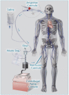
Figure 4. AngioVac system. AngioVac system (courtesy of Angiodynamics, Latham, NY).
Figure 5
Figure 5. Tricuspid valve methicillin‐sensitive Staphylococcus aureus MSSA IE and pacemaker‐lead Staphylococcus IE cases treated with AngioVac system.
A through D, A 28‐year old female with a history of congenital long QT syndrome and prophylactic implantable cardioverter defibrillator implantation at the age of 14. She underwent several generator replacements and lead revisions, with the last one occurring 1 month prior to her admission. She was admitted due to fever and chills, a new systolic murmur and splenomegaly on physical examination. Blood cultures were positive for coagulase negative staphylococcus. TTE was nondiagnostic; however, two‐dimensional (2D) (A) and three‐dimensional (3D) (B) TEE showed a large adherent mass on the pacemaker lead (blue arrow). The patient underwent lead extraction; however, following the procedure she developed pulmonary emboli. Repeated TEE showed a large residual tricuspid vegetation (C, red arrow). Therefore, a percutaneous vacuum‐assisted aspiration device was used to successfully remove the residual tricuspid vegetation. Following the aspiration procedure, no vegetations were observed by TEE (D), and the patient's infection completely resolved. E and F, A 33‐year‐old female with history of IVDU was admitted due to fever. Blood cultures were positive for methicillin‐sensitive Staphylococcus aureus (MSSA), which did not respond to oxacillin treatment. TEE revealed a large tricuspid valve vegetation (E, yellow arrow) with significant tricuspid regurgitation. Her hospital course was complicated by septic pulmonary emboli and hemoptysis. She was considered high risk for surgery and therefore underwent a successful percutaneous vacuum‐assisted aspiration of her tricuspid vegetation. On follow‐up TEE a few days after there were no evidence of any tricuspid vegetation and only mild tricuspid regurgitation (F). Her condition gradually improved, and she was discharged home. Nine months later, her follow‐up TTE showed no evidence of vegetations. 3D indicates three‐dimensional; AV, aortic valve; IVDU, intravenous drug user; LA, left atrium; LV, left ventricle; RA, right atrium; RV, right ventricle; TEE, transesophageal echocardiography; and TTE, transthoracic echocardiography.

References

    1. Geller SA. Infective endocarditis: a history of the development of its understanding. Autops Case Rep. 2013;3:5–12. - PMC - PubMed
    1. Cahill TJ, Prendergast BD. Infective endocarditis. Lancet. 2016;387:882–893. - PubMed
    1. Pant S, Patel NJ, Deshmukh A, Golwala H, Patel N, Badheka A, Hirsch GA, Mehta JL. Trends in infective endocarditis incidence, microbiology, and valve replacement in the United States from 2000 to 2011. J Am Coll Cardiol. 2015;65:2070–2076. - PubMed
    1. Sambola A, Fernandez‐Hidalgo N, Almirante B, Roca I, Gonzalez‐Alujas T, Serra B, Pahissa A, Garcia‐Dorado D, Tornos P. Sex differences in native‐valve infective endocarditis in a single tertiary‐care hospital. Am J Cardiol. 2010;106:92–98. - PubMed
    1. Correa de Sa DD, Tleyjeh IM, Anavekar NS, Schultz JC, Thomas JM, Lahr BD, Bachuwar A, Pazdernik M, Steckelberg JM, Wilson WR, et al. Epidemiological trends of infective endocarditis: a population‐based study in Olmsted County, Minnesota. Mayo Clin Proc. 2010;85:422–426. - PMC - PubMed

MeSH terms

Substances