. 2020 Oct;44(4):1415-1424.
doi: 10.3892/or.2020.7700. Epub 2020 Jul 21.

Sensitivity of allyl isothiocyanate to induce apoptosis via ER stress and the mitochondrial pathway upon ROS production in colorectal adenocarcinoma cells

Affiliations

Sensitivity of allyl isothiocyanate to induce apoptosis via ER stress and the mitochondrial pathway upon ROS production in colorectal adenocarcinoma cells

Jo-Hua Chiang et al. Oncol Rep. 2020 Oct.

Abstract

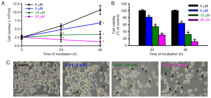
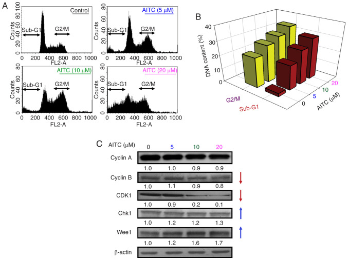
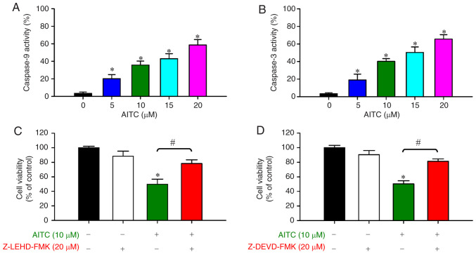
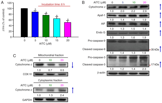
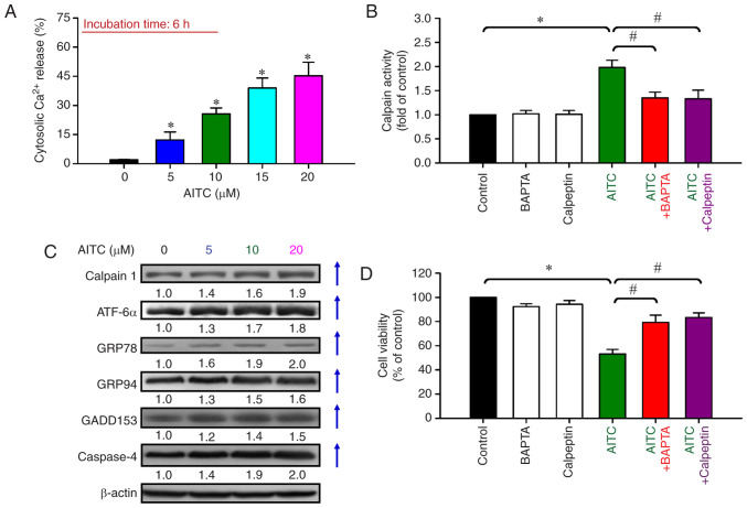
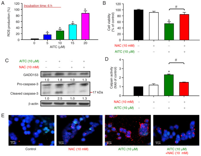
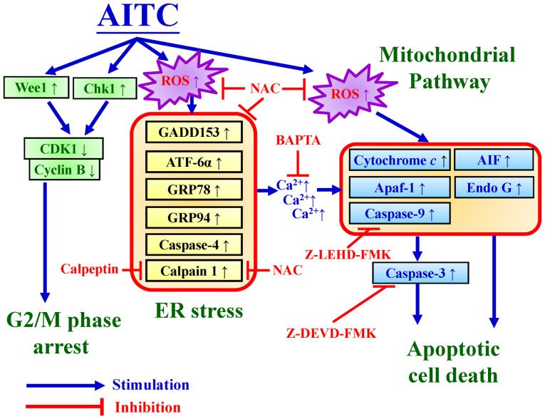
Allyl isothiocyanate (AITC), a bioactive phytochemical compound that is a constituent of dietary cruciferous vegetables, possesses promising chemopreventive and anticancer effects. However, reports of AITC exerting antitumor effects on apoptosis induction of colorectal cancer (CRC) cells in vitro are not well elucidated. The present study focused on the functional mechanism of the endoplasmic reticulum (ER) stress‑based apoptotic machinery induced by AITC in human colorectal cancer HT‑29 cells. Our results indicated that AITC decreased cell growth and number, reduced viability, and facilitated morphological changes of apoptotic cell death. DNA analysis by flow cytometry showed G2/M phase arrest, and alterations in the modulated protein levels caused by AITC were detected via western blot analysis. AITC also triggered vital intrinsic apoptotic factors (caspase‑9/caspase‑3 activity), disrupted mitochondrial membrane potential, and stimulated mitochondrial‑related apoptotic molecules (e.g., cytochrome c, apoptotic protease activating factor 1, apoptosis‑inducing factor, and endonuclease G). Additionally, AITC prompted induced cytosolic Ca2+ release and Ca2+‑dependent ER stress‑related signals, such as calpain 1, activating transcription factor 6α, glucose‑regulated proteins 78 and 94, growth arrest‑ and DNA damage‑inducible protein 153 (GADD153), and caspase‑4. The level of reactive oxygen species (ROS) production was found to induce the hallmark of ER stress GADD153, proapoptotic marker caspase‑3, and calpain activity after AITC treatment. Our findings showed for the first time that AITC induced G2/M phase arrest and apoptotic death via ROS‑based ER stress and the intrinsic pathway (mitochondrial‑dependent) in HT‑29 cells. Overall, AITC may exert an epigenetic effect and is a potential bioactive compound for CRC treatment.

Keywords: allyl isothiocyanate; colon cancer cells; apoptosis; ER stress; mitochondria; ROS production.


Figures

Figure 1.
Figure 1.
Effects of AITC on the number, viability, and morphology of HT-29 cells. The cells were incubated with AITC (0, 5, 10 and 20 µM) for 24 and 48 h. (A) Cell counting assay was performed via the trypan blue exclusion method. (B) The viability of HT-29 cells was detected using MTT assay. Each point represents mean ± SD of three experiments; *P<0.05 vs. the control (Dunnett's post hoc test after ANOVA). (C) After treatment with AITC for 24 h, cells in each well were visualized and photographed using phase-contrast microscopy at ×200 magnification. Representative images are shown. The arrows (↑) indicate apoptotic cells. Scale bar, 15 µm. AITC, allyl isothiocyanate.
Figure 2.
Figure 2.
Effects of AITC on cell cycle progression and G2/M phase-regulated protein levels of HT-29 cells. The cells were exposed to various concentrations (0, 5, 10 and 20 µM) of AITC for 24 h. (A) Representative images of the cell cycle distribution, as determined using flow cytometric analysis. (B) Quantification of G2/M phase and sub-G1 population. (C) The total protein in the cell extracts was subjected to western blot to detect G2/M progression-associated proteins (cyclin A, cyclin B, CDK1, Chk1 and Wee1). All blots were normalized to β-actin to ensure the same amount of loading of each sample across all samples. AITC, allyl isothiocyanate; CDK1, cyclin-dependent kinase 1; Chk1, checkpoint kinase 1.
Figure 3.
Figure 3.
Effects of AITC on caspase-9 and caspase-3 activities of HT-29 cells. Cells were treated with AITC (0, 5, 10, 15 and 20 µM) for 24 h. Cell lysates were harvested, and (A) caspase-9 and (B) caspase-3 activities were determined, as described in Materials and methods. *P <0.05 vs. the control; statistical significance was determined by ANOVA (Dunnett's test). The cells were incubated with 10 µM AITC for 24 h after pretreatment with or without (C) Z-LEDH-FMK (20 µM, a caspase-9 inhibitor) and (D) Z-DEVD-FMK (20 µM, a caspase-3 inhibitor) for 2 h to monitor cell viability via MTT assay. *P<0.05 vs. the control and #P<0.05 vs. AITC-treated only cells; statistically significant differences were determined by ANOVA (Tukey's test). Each point represents the mean ± SD of three experiments. AITC, allyl isothiocyanate.
Figure 4.
Figure 4.
Effects of AITC on the mitochondrial-dependent apoptotic pathway of HT-29 cells. (A) The cells were incubated with AITC (0, 5, 10, 15 and 20 µM) for 6 h and then harvested to examine the level of ΔΨm via DiOC6(3) and flow cytometry. Each point represents the mean ± SD of three experiments; *P<0.05 vs. the control (Dunnett's test after ANOVA). (B) Cells were exposed to the indicated concentrations of AITC for 24 h, and the cell fraction was prepared and analyzed via western blot analysis to estimate the levels of cytochrome c, Apaf-1, AIF, Endo G, caspase-9 and caspase-3 protein expression. All blots were normalized to β-actin to ensure the same amount of loading of each sample across all samples. (C) The mitochondrial (top) and cytoplasmic (bottom) fractions were prepared to detect cytochrome c trafficking via western blot analysis. COX IV and GAPDH were analyzed to ensure the same amount of loading. AITC, allyl isothiocyanate; Apaf-1, apoptotic protease activating factor 1; AIF, apoptosis-inducing factor; Endo G, endonuclease G.
Figure 5.
Figure 5.
Effects of AITC on cytosolic Ca2+ and ER stress signaling of HT-29 cells. (A) Cells were treated with AITC (0, 5, 10, 15 and 20 µM) for 6 h, and the level of cytosolic Ca2+ was assessed via fluo-3/AM and flow cytometry. *P<0.05 vs. the control; statistical significance was determined by ANOVA (Dunnett's test). (B) The cells were pretreated with or without 5 µM BAPTA/AM or 20 µM calpain for 2 h and then exposed to 10 µM AITC for 24 h. The cells were harvested and examined for calpain activity. (C) The protein levels of calpain 1, ATF-6α, GRP78, GRP94, GADD153, and caspase-4 were determined using western blot analysis. All blots were normalized to β-actin to ensure the same amount of loading of each sample across all samples. (D) Cell viability was detected using MTT assay. *P<0.05 vs. the control and #P<0.05 vs. AITC-treated only cells; statistically significant differences were determined by ANOVA (Tukey's test). Each point represents the mean ± SD of three experiments; AITC, allyl isothiocyanate; ER, endoplasmic reticulum; ATF-6α, activating transcription factor 6α; GRP78, 78 kDa glucose-regulated protein; GRP94, 94 kDa glucose-regulated protein; GADD153, growth arrest- and DNA damage-inducible protein 153.
Figure 6.
Figure 6.
Effects of AITC on ROS production, GADD153 (the hallmark of ER stress), and caspase-3 (an apoptotic marker) of HT-29 cells. (A) Cells were incubated with or without 5, 10, 15 and 20 µM of AITC for 6 h, and the level of ROS generation was measured using ROS indicator H2DCFDA and flow cytometry. *P<0.05 vs. the control; statistical significance was determined by ANOVA (Dunnett's test). (B) The cells were preincubated with or without 10 mM NAC for 2 h prior to 10 µM AITC exposure for 24 h. Cell viability was analyzed using MTT assay. (C) After AITC exposure for 24 h, the cells were collected and the protein levels of GADD153 and caspase-3 were estimated via western blot analysis. All blots were normalized to β-actin to ensure the same amount of loading of each sample across all samples. (D) After pretreatment with or without 10 mM NAC for 2 h, the cells were exposed to 10 µM AITC exposure for 24 h and then monitored for calpain activity. *P<0.05 vs. the control and #P<0.05 vs. AITC-treated only cells; statistically significant differences were determined by ANOVA (Tukey's test). Each point represents mean ± SD of three experiments; (E) The protein expression of calpain 1 was visualized, and the photomicrographs were analyzed via immunofluorescence staining using confocal microscopy of calpain 1 (red, PE-labeled) and nuclei (blue, DAPI-stained). Scale bar, 20 µm. AITC, allyl isothiocyanate; ROS, reactive oxygen species; ER, endoplasmic reticulum; GADD153, growth arrest- and DNA damage-inducible protein 153.
Figure 7.
Figure 7.
Schematic diagram of the interplay among G2/M phase arrest, ER stress, and mitochondrial-dependent apoptosis in human colorectal adenocarcinoma HT-29 cells. Blockage of G2/M phase by AITC occurs by modulating Wee1, Chk1, and CDK1/cyclin B signal molecules. ROS production induced by AITC fully facilitates ER stress and mitochondrial apoptotic mechanisms. As a result, ER stress under oxidative stress (ROS production) conditions may contribute to AITC-provoked apoptotic machinery in HT-29 cells. AITC, allyl isothiocyanate; ROS, reactive oxygen species; NAC, N-acetylcysteine; CDK1, cyclin-dependent kinase 1; Chk1, checkpoint kinase 1; GADD153, growth arrest- and DNA damage-inducible protein 153; Apaf-1, apoptotic protease activating factor 1; AIF, apoptosis-inducing factor; Endo G, endonuclease G; ATF-6α, activating transcription factor 6α; GRP78, 78 kDa glucose-regulated protein; GRP94, 94 kDa glucose-regulated protein.

References

    1. Ferlay J, Colombet M, Soerjomataram I, Dyba T, Randi G, Bettio M, Gavin A, Visser O, Bray F. Cancer incidence and mortality patterns in Europe: Estimates for 40 countries and 25 major cancers in 2018. Eur J Cancer. 2018;103:356–387. doi: 10.1016/j.ejca.2018.07.005. - DOI - PubMed
    1. Shen W, Jin Z, Tong X, Wang H, Zhuang L, Lu X, Wu S. TRIM14 promotes cell proliferation and inhibits apoptosis by suppressing PTEN in colorectal cancer. Cancer Manag Res. 2019;11:5725–5735. doi: 10.2147/CMAR.S210782. - DOI - PMC - PubMed
    1. Ribeiro Gomes J, Belotto M, D'Alpino Peixoto R. The role of surgery for unusual sites of metastases from colorectal cancer: A review of the literature. Eur J Surg Oncol. 2017;43:15–19. doi: 10.1016/j.ejso.2016.05.019. - DOI - PubMed
    1. Cheng X, Feng H, Wu H, Jin Z, Shen X, Kuang J, Huo Z, Chen X, Gao H, Ye F, et al. Targeting autophagy enhances apatinib-induced apoptosis via endoplasmic reticulum stress for human colorectal cancer. Cancer Lett. 2018;431:105–114. doi: 10.1016/j.canlet.2018.05.046. - DOI - PubMed
    1. Afrin S, Giampieri F, Gasparrini M, Forbes-Hernández TY, Cianciosi D, Reboredo-Rodriguez P, Zhang J, Manna PP, Daglia M, Atanasov AG, Battino M. Dietary phytochemicals in colorectal cancer prevention and treatment: A focus on the molecular mechanisms involved. Biotechnol Adv. 2020;38:107322. doi: 10.1016/j.biotechadv.2018.11.011. - DOI - PubMed

MeSH terms