

99mTc-Leukocyte Scintigraphy Revealed Viral Pulmonary Infection in a COVID-19 Patient
- PMID: 32701817
- PMCID: PMC7473795
- DOI: 10.1097/RLU.0000000000003219
99mTc-Leukocyte Scintigraphy Revealed Viral Pulmonary Infection in a COVID-19 Patient
Abstract
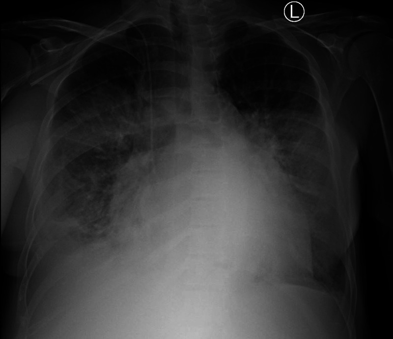
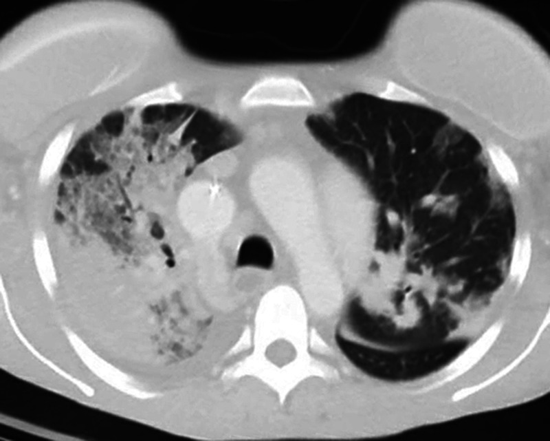
Tc-leukocyte scintigraphy was performed on a 40-year-old woman with spiking fevers. A focus of intense uptake in the right upper thorax was identified, concerning for infection along the central line in the superior vena cava. Additionally, heterogeneously increased uptake in both lungs was noted, which suggested pulmonary infection. CT images of the chest showed patchy ground-glass changes in both lungs and a large consolidation in the right lower lobe, which were consistent with changes for COVID-19 (coronavirus disease 2019). Severe acute respiratory syndrome coronavirus 2 RNA test was positive. This case demonstrates that leukocyte uptake in bilateral lungs could reveal viral pulmonary infection in COVID-19.
Conflict of interest statement
Conflicts of interest and sources of funding: none declared.
Figures

Similar articles
-
Chest CT imaging characteristics of COVID-19 pneumonia in preschool children: a retrospective study.BMC Pediatr. 2020 May 18;20(1):227. doi: 10.1186/s12887-020-02140-7. BMC Pediatr. 2020. PMID: 32423435 Free PMC article.
-
Clinical and computed tomographic imaging features of novel coronavirus pneumonia caused by SARS-CoV-2.J Infect. 2020 Apr;80(4):394-400. doi: 10.1016/j.jinf.2020.02.017. Epub 2020 Feb 25. J Infect. 2020. PMID: 32109443 Free PMC article.
-
A Case of Coronavirus Infection Incidentally Found on FDG PET/CT Scan.Clin Nucl Med. 2020 Jul;45(7):e303-e304. doi: 10.1097/RLU.0000000000003084. Clin Nucl Med. 2020. PMID: 32433168 Free PMC article.
-
Diagnostic value and key features of computed tomography in Coronavirus Disease 2019.Emerg Microbes Infect. 2020 Dec;9(1):787-793. doi: 10.1080/22221751.2020.1750307. Emerg Microbes Infect. 2020. PMID: 32241244 Free PMC article. Review.
-
Computed Tomography Features of Coronavirus Disease 2019 (COVID-19): A Review for Radiologists.J Thorac Imaging. 2020 Jul;35(4):211-218. doi: 10.1097/RTI.0000000000000527. J Thorac Imaging. 2020. PMID: 32427651 Review.
Cited by
-
Diagnostic, Prognostic, and Therapeutic Use of Radiopharmaceuticals in the Context of SARS-CoV-2.ACS Pharmacol Transl Sci. 2021 Jan 15;4(1):1-7. doi: 10.1021/acsptsci.0c00186. eCollection 2021 Feb 12. ACS Pharmacol Transl Sci. 2021. PMID: 33615159 Free PMC article. Review.
-
Radiological and Imaging Evidence in the Diagnosis and Management of Microbial Infections: An Update.Cureus. 2023 Nov 13;15(11):e48756. doi: 10.7759/cureus.48756. eCollection 2023 Nov. Cureus. 2023. PMID: 38094521 Free PMC article. Review.
-
Nuclear Medicine in Times of COVID-19: How Radiopharmaceuticals Could Help to Fight the Current and Future Pandemics.Pharmaceutics. 2020 Dec 21;12(12):1247. doi: 10.3390/pharmaceutics12121247. Pharmaceutics. 2020. PMID: 33371500 Free PMC article. Review.
-
99mTc-Vitamin C SPECT/CT imaging in SARS-CoV-2 associated pneumonia.Nucl Med Rev Cent East Eur. 2022;25(2):127-128. doi: 10.5603/NMR.a2022.0026. Epub 2022 Jun 14. Nucl Med Rev Cent East Eur. 2022. PMID: 35699590 Free PMC article.
-
Lymphopenia in patients affected by SARS-CoV-2 infection is caused by margination of lymphocytes in large bowel: an [18F]FDG PET/CT study.Eur J Nucl Med Mol Imaging. 2022 Aug;49(10):3419-3429. doi: 10.1007/s00259-022-05801-0. Epub 2022 Apr 29. Eur J Nucl Med Mol Imaging. 2022. PMID: 35486145 Free PMC article.
References
-
- Palestro CJ Brown ML Forstrom LA, et al. . Society of Nuclear Medicine procedure guideline for 99mTc-exametazime (HMPAO)–labeled leukocyte scintigraphy for suspected infection/inflammation, version 3.0. 2004. Available at: http://interactive.snm.org/docs/HMPAO_v3.pdf 11.
-
- Love C, Palestro CJ. Radionuclide imaging of infection. J Nucl Med Technol. 2004;32:47–57. - PubMed
-
- Czernin J Fanti S Meyer P, et al. . Nuclear medicine operations in the times of COVID-19: strategies, precautions, and experiences. J Nucl Med. 2020;61:626–629. - PubMed
Publication types
MeSH terms
Substances
LinkOut - more resources
Full Text Sources
Medical
