. 2020 Dec;99(13):1478-1485.
doi: 10.1177/0022034520941837. Epub 2020 Jul 23.

Transcriptomic Classification of Neurons Innervating Teeth

Affiliations

Transcriptomic Classification of Neurons Innervating Teeth

J J Emrick et al. J Dent Res. 2020 Dec.

Abstract

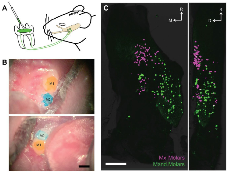
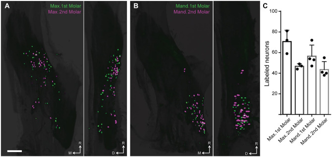
Toothache is a common painful consequence of damage to the teeth, particularly when coupled to infection. Clinical restoration of tooth integrity, sometimes involving physical and chemical sterilization of the tooth with nerve fiber ablation (i.e., endodontic therapy), generally alleviates pain and allows long-lasting dental function. These observations raise questions regarding the biological role of tooth-innervating fibers. Here, we determined the transcriptomic diversity of the sensory neurons that can be retrogradely labeled from mouse molar teeth. Our results demonstrate that individual molars are each targeted by a dedicated population of about 50 specialized trigeminal neurons. Transcriptomic profiling identifies the majority of these as expressing markers of fast-conducting neurons, with about two-thirds containing nociceptive markers. Our data provide a new view of dental innervation, extending previous reports that used candidate gene approaches. Importantly, almost all retrogradely labeled neurons, including nociceptors, express the recently characterized mechanosensor Piezo2, an ion channel that endows cells with sensitivity to gentle touch. Intriguingly, about a quarter of the labeled neurons do not appear to be nociceptors, perhaps insinuating a role for them in discriminative touch. We hypothesize that dental neurons are capable of providing mechanosensitive information to drive rapid behavioral responses and protect teeth from damage. Damage to the teeth and exposure of the large population of molar nociceptors may trigger prolonged or abnormal activation driving toothache. Future studies examining the responses of these transcriptomically defined classes of neurons will help define their significance in oral sensation.

Keywords: gene expression; mechanotransduction; neuroscience/neurobiology; pain; pulp biology; receptors.


Conflict of interest statement

The authors declare no potential conflicts of interest with respect to the authorship and/or publication of this article.

Figures

Figure 1.
Figure 1.
Retrograde labeling of the trigeminal sensory neurons reveals rich and selective innervation of molar teeth. (A) Schematic depicting our labeling paradigm: briefly, we prepared shallow surgical cavitations in the mouse molars, applied retrograde tracer, and observed robust labeling in somata of trigeminal sensory neurons from 16 to 72 h after labeling. (B) Images of maxillary (top) and mandibular molars (bottom) after application of distinct retrograde tracers CTB-488 (yellow) and CTB-647 (blue) and placement of dental composite. Scale bar: 500 μm. CTB, cholera toxin B-subunit. M1 and M2 denote first and second molar within each arch. (C) Representative light sheet microscopy of an optically cleared right trigeminal ganglion with sensory neurons labeled from application of spectrally distinct CTB conjugates to ipsilateral maxillary (magenta) and mandibular (green) arches (the first and second molars in each quadrant are labeled). Two-dimensional maximum projection images of the whole ganglion from the dorsal (left panel) and lateral (right panel) views with rostral orientation toward the top. Scale bar: 500 μm. D, dorsal; M, medial; R, rostral. See Appendix Movie 1 for 3-dimensional reconstruction of these data.
Figure 2.
Figure 2.
Adjacent mouse molars are targeted by distinct populations of sensory neurons. (A, B) Representative collapsed images (maximum projection light sheet) of optically cleared right trigeminal ganglia with sensory neurons labeled from application of spectrally distinct CTB conjugates to first (green) and second (magenta) molars. View of the whole ganglion from the dorsal (left panel) and lateral (right panel) views with the rostral orientation toward the top. (a) Maxillary and (b) mandibular teeth were labeled in different animals. Scale bar: 500 μm. CTB, cholera toxin B-subunit; D, dorsal; M, medial; R, rostral. (C) Bar graph representing the number of trigeminal sensory neurons labeled following application of single tracer to each tooth. Data are mean ± SEM from 3 mice for maxillary teeth and 4 mice from mandibular teeth. See Appendix Movies 2 and 3 for 3-dimensional reconstruction of these data.
Figure 3.
Figure 3.
Multigene in situ hybridization (ISH) reveals that CTB-labeled molar neurons are dominated by 2 transcriptomic classes of large A-type sensory neurons. (A) Representative images from retrograde labeling of molar neurons and multigene ISH for a single section of a trigeminal ganglion. CTB–Alexa 555 was applied to the maxillary first and second molars; retrograde-labeled neurons were imaged (red); and the section was subjected to 3 rounds of ISH (green). Genes (left to right, beginning in the top row): S100 calcium-binding protein B (S100b), calcitonin gene-related peptide (CGRP; Calca), FXYD domain-containing ion transport regulator 2 (Fxyd2), natriuretic polypeptide B (Nppb), transmembrane protein 233 (Tmem233), mas-related G protein–coupled receptor member D (Mrgprd); transient receptor potential cation channel, subfamily A, member 1 (Trpa1); and transient receptor potential cation channel, subfamily M, member 8 (Trpm8). To aid identification of molar neurons, the retrograde labeling was overlaid on individual ISH images. Scale bar: 100 μm. CTB, cholera toxin B-subunit. Transcriptomic class of 401 molar neurons from 6 mice was determined per an automated UNet-based algorithm. Data are from 55 fields. (B) Stacked bar charts comparing the transcriptomic profile of molar neurons (left) with a general population of trigeminal sensory neurons from the whole ganglion (right). (C) Algorithm criteria for transcriptomic class assignment.
Figure 4.
Figure 4.
WGA and CTB tracing reveal similar comprehensive labeling and classification of molar neurons. (A) Collapsed image (maximum projection light sheet) of optically cleared right trigeminal ganglia with sensory neurons labeled from application of WGA–Alexa 488 (green) and CTB–Alexa 647 (magenta) to the maxillary first molar. Dorsal view with rostral orientation toward the left. Scale bar: 500 μm. L, lateral; R, rostral. (B) Panels highlight extensive overlap of CTB and WGA labeling. The magnified field is indicated by a white rectangle in panel A. Scale bar: 100 μm. (C) Images are shown for comparison from an optically cleared trigeminal ganglion with sensory neurons labeled by injection of spectrally distinct WGA and CTB tracers into the attached gingiva covering the hard palate. Note the larger number of small-diameter cells and the lower degree of overlap between the labels. Scale as shown in panel B. Similar results were obtained for these types of colabeling in 2 animals for each site of application. (D) Pie charts representing the transcriptomic classification of WGA- and CTB-labeled neurons innervating the palate (1635 neurons, 2 groups of 3 mice, 55 fields) and first and second maxillary molars (812 neurons, 4 groups of 3 mice, 97 fields) highlight that, whereas many classes of sensory neuron target the palate, the molar teeth are innervated primarily by C4 and C6 cells. (E) For cells targeting the molars (black solid bars) and the palate (gray open bars), fold enrichment of each class of neurons was calculated relative to prevalence in the whole ganglion (see Fig. 3B). Bar graph shows the mean + SEM (n = 4 groups, molars; 2 groups, palate) and the individual results from WGA (red dots) and CTB labeling (green dots). All classes other than C4 and C6 innervate the teeth less frequently than what would be expected (dashed line). See Appendix Table 2 for statistical analysis. (F) Distribution of diameters for CTB- and WGA-labeled cells targeting the molars (black solid bars) and the palate (gray open bars) as quantitated in tissue sections. CTB, cholera toxin B-subunit; WGA, wheat germ agglutinin.
Figure 5.
Figure 5.
Molars are innervated by distinct populations of large-diameter sensory neurons involved in nociception and discriminative touch. (A) Representative images are shown from retrograde tracing (WGA) and 6 rounds of multigene in situ hybridization for a single section of a trigeminal ganglion. Expression patterns of a panel of genes in molar neurons labeled by application of WGA–Texas red to the maxillary first and second molars allowed expanded classification by transcriptomic profile. Fluorescence microscopy images show a region of the ganglion with retrogradely labeled molar neurons (WGA). Red circles outline neurons labeled by WGA, allowing visual identification of their gene expression pattern. WGA staining and the genes used for 8-probe classification are shown in the top 2 rows. Other genes that map to subsets of C4 and C6 cells in single nuclear (sn) RNA sequencing (Nguyen et al. 2019) were examined (see Appendix Table 1 for details of their expression and properties). Genes were as follows: 5-hydroxytryptamine (serotonin) receptor 3A (Htr3a); transient receptor potential cation channel, subfamily V, member 1 (Trpv1); sodium channel, voltage-gated, type X, alpha-Nav1.8 (Scn10a); teashirt zinc finger family member 2 (Tshz2); neurofilament, heavy polypeptide-NF200 (Nefh); piezo-type mechanosensitive ion channel component 2 (Piezo2); protocadherin 7 (Pcdh7); bone morphogenetic protein receptor 1b (Bmpr1b); netrin G1 (Ntng1); and calretinin (Calb2). Data are from 44 fields. Scale bar: 100 μm. (B) Venn diagram indicates number and percentage of neurons expressing selected genes in the S100b-positive labeled cells (87%; 580 of 668 total molar neurons). Note that a fraction of the C6 cells were positive for Trpv1 and that Htr3a and Scn10a, genes involved in nociception, labeled large groups of C4 molar neurons. (C) Venn diagram depicting the expression of Piezo2 in neuron populations found in panel B. CTB, cholera toxin B-subunit; WGA, wheat germ agglutinin.

References

    1. Abrahamsen B, Zhao J, Asante CO, Cendan CM, Marsh S, Martinez-Barbera JP, Nassar MA, Dickenson AH, Wood JN. 2008. The cell and molecular basis of mechanical, cold, and inflammatory pain. Science. 321(5889):702–705. - PubMed
    1. Chaudhary P, Martenson ME, Baumann TK. 2001. Vanilloid receptor expression and capsaicin excitation of rat dental primary afferent neurons. J Dent Res. 80(6):1518–1523. - PubMed
    1. Chesler AT, Szczot M, Bharucha-Goebel D, Čeko M, Donkervoort S, Laubacher C, Hayes LH, Alter K, Zampieri C, Stanley C, et al. 2016. The role of PIEZO2 in human mechanosensation. N Engl J Med. 375(14):1355–1364. - PMC - PubMed
    1. Chung MK, Jue SS, Dong X. 2012. Projection of non-peptidergic afferents to mouse tooth pulp. J Dent Res. 91(8):777–782. - PMC - PubMed
    1. Chung MK, Lee J, Duraes G, Ro JY. 2011. Lipopolysaccharide-induced pulpitis up-regulates TRPV1 in trigeminal ganglia. J Dent Res. 90(9):1103–1107. - PMC - PubMed

Publication types

LinkOut - more resources