. 2020 Jul 23;18(1):115.
doi: 10.1186/s12964-020-00584-z.

AMPK activation overcomes anti-EGFR antibody resistance induced by KRAS mutation in colorectal cancer

Affiliations

AMPK activation overcomes anti-EGFR antibody resistance induced by KRAS mutation in colorectal cancer

Hua Ye et al. Cell Commun Signal. .

Abstract

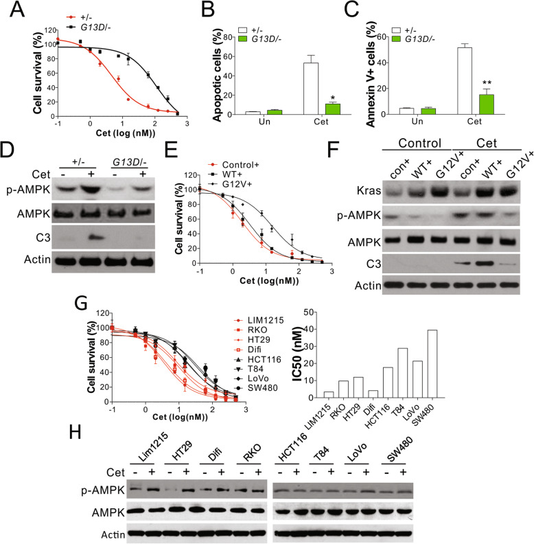
Background: Colorectal cancer (CRC) is associated with resistance to anti-epidermal growth factor receptor (EGFR) antibodies (both acquired and intrinsic), owing to the amplification or mutation of the KRAS oncogene. However, the mechanism underlying this resistance is incompletely understood.

Methods: DLD1 cells with WT (+/-) or KRAS G13D mutant allele were treated with different concentrations of Cetuximab (Cet) or panitumumab (Pab) to study the mechanism underlying the KRAS mutation-induced resistance to anti-EGFR antibodies. The function of AMPK in KRAS mutation-induced resistance to anti-EGFR antibodies in CRC cells, and the regulatory role of Bcl-2 family proteins in DLD1 cells with WT or mutated KRAS upon AMPK activation were investigated. In addition, xenograft tumor models with the nude mouse using DLD1 cells with WT or mutated KRAS were established to examine the effects of AMPK activation on KRAS mutation-mediated anti-EGFR antibody resistance.

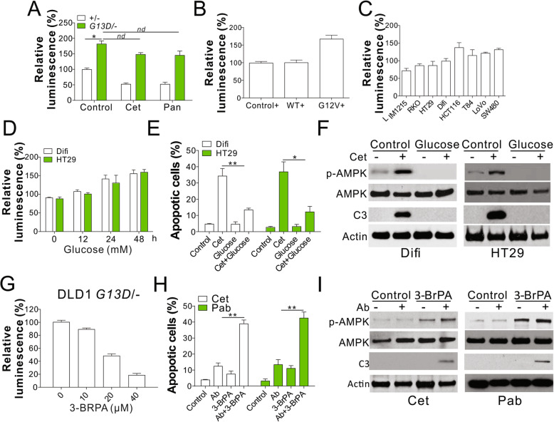
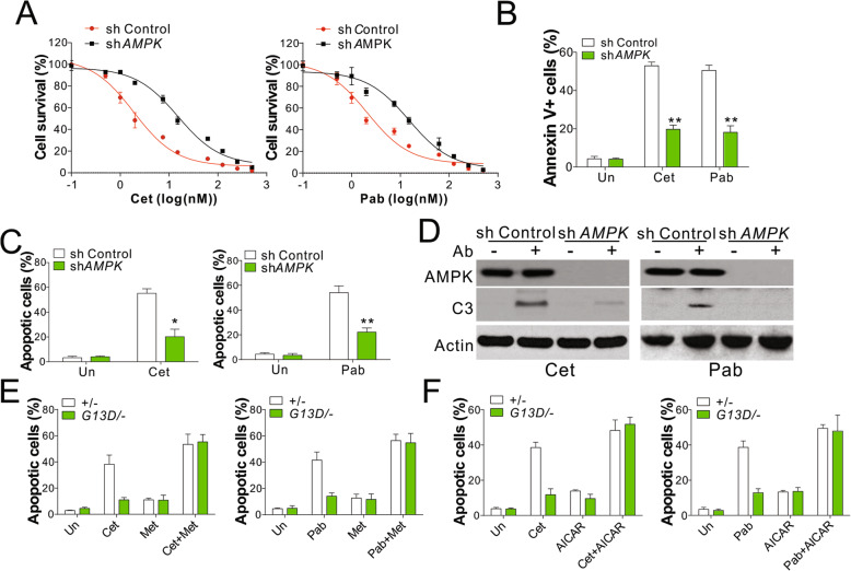
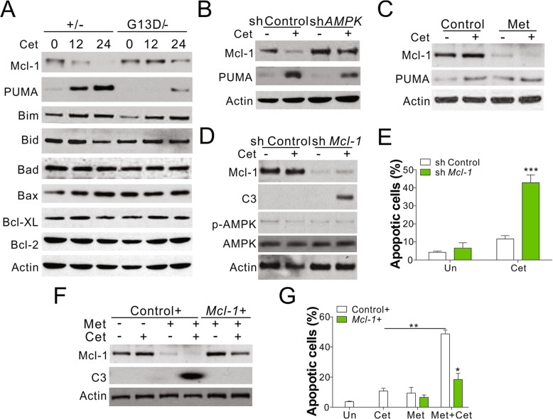
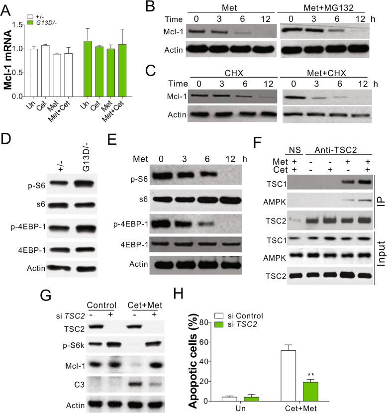
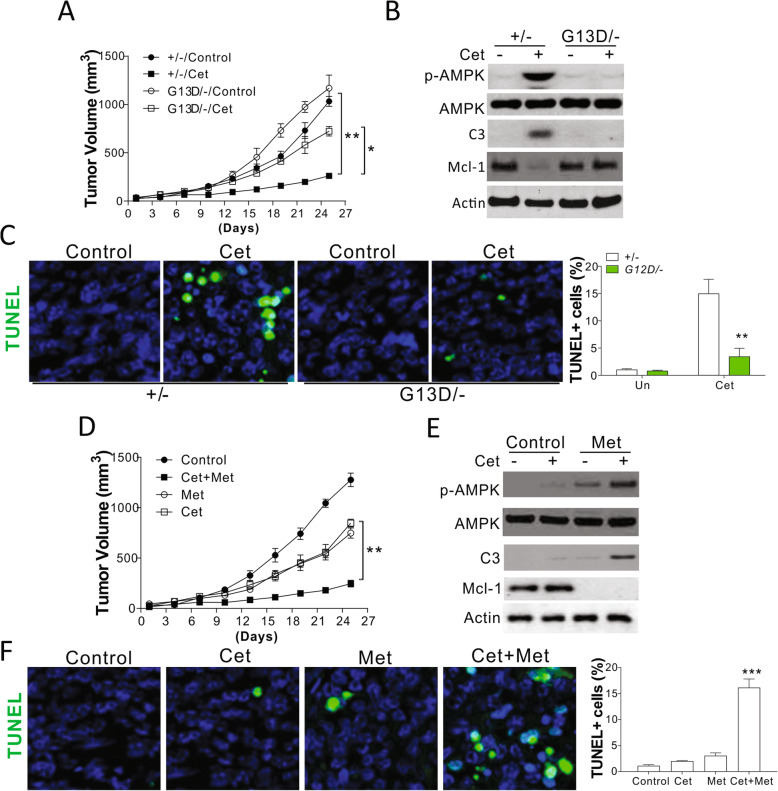
Results: Higher levels of AMPK activity in CRC cells with wild-type KRAS treated with anti-EGFR antibody resulted in apoptosis induction. In contrast, CRC cells with mutated KRAS showed lower AMP-activated protein kinase (AMPK) activity and decreased sensitivity to the inhibitory effect of anti-EGFR antibody. CRC cells with mutated KRAS showed high levels of glycolysis and produced an excessive amount of ATP, which suppressed AMPK activation. The knockdown of AMPK expression in CRC cells with WT KRAS produced similar effects to those observed in cells with mutated KRAS and decreased their sensitivity to cetuximab. On the contrary, the activation of AMPK by metformin (Met) or 5-aminoimidazole-4-carboxamide ribonucleotide (AICAR) could overcome the KRAS-induced resistance to the anti-EGFR antibody in vivo and in vitro. The activation of AMPK resulted in the inhibition of myeloid cell leukemia 1 (Mcl-1) translation through the suppression of the mammalian target of rapamycin (mTOR) pathway.

Conclusion: The results established herein indicate that targeting AMPK is a potentially promising and effective CRC treatment strategy. Video abstract.

Keywords: AMPK; CRC; EGFR.


Conflict of interest statement

The authors declare that they have no competing interests.

Figures

Fig. 1
Fig. 1
Phosphorylation of AMPK was suppressed by Kras mutation in CRC cells. a MTS analysis of DLD1 WT (+/−) and Kras mutation (G13D/−) cells treated with cetuximab (Cet) at the indicated doses for 48 h. b Apoptosis in DLD1 WT (+/−) and Kras mutation (G13D/−) cells treated with 5 nM Cet for 48 h was analyzed by nuclear staining with Hoechst 33258. c Apoptosis in DLD1 WT (+/−) and Kras mutation (G13D/−) cells treated with 5 nM Cet for 48 h was analyzed by Annexin V staining followed by flow cytometry. d Western blot of p-AMPK, AMPK, and Caspase-3 (C3) in the cells treated as in (b). e DLD1 WT (+/−) cells stably expressing control, Kras WT, or Kras mutant (G12V) by retrovirus transfection were treated with Cet at indicated doses for 48 h. The cell viability was analyzed by MTS assay. f DLD1 WT (+/−) cells stably expressing control, Kras WT, or Kras mutant (G12V) by retrovirus transfection were treated with 5 nM Cet for 48 h. The expression of indicated proteins was analyzed western blot. g MTs analysis of indicated cells treated with Cet at indicated doses for 48 h. The IC50 was calculated and plotted in the right panel. h The expression of p-AMPK, AMPK in the indicated cells treated with 5 nM Cet. Each experiment was repeated for 3 times. *, p < 0.05; **, p < 0.01
Fig. 2
Fig. 2
Kras mutation suppressed the AMPK activation by glycolysis. a The cellular ATP level of DLD1 WT and Kras mutation cells was analyzed by luminescence assay. b The cellular ATP level in DLD1 WT (+/−) cells stably expressing control, Kras WT, or Kras mutant (G12V) by retrovirus transfection (c) The cellular ATP level in the indicated cell lines. d The cellular ATP level of HT29 and Difi cells cultured in media containing 20 mM glucose at indicated time. e HT29 and Difi cells cultured in the media with or without 20 mM glucose were treated with Cet (15 nM for HT29; 5 nM for Difi) for 48 h. The apoptosis was analyzed by Hoechst 33258 staining. f Western blot of indicated proteins in HT29 and Difi cells treated as in (e). g The cellular ATP level of DLD1 Kras mutated treated with 10 μM 3-BrPA for indicated time points. h The apoptosis of DLD1 Kras mutated cells treated with 10 μM 3-BrPA in combined with 5 nM Cet or 10 nM panitumumab (Pab). i Western blot of indicated proteins in DLD1 Kras mutated cells treated as in (h). Each experiment was repeated for 3 times. nd, p > 0.05; *, p < 0.05; **, p < 0.01
Fig. 3
Fig. 3
Activation of AMPK reversed the anti-EGFR antibodies resistance induced by Kras mutation. a MTS analysis of DLD1 WT (+/−) transfected with control or AMPK shRNA and treated with Cet (left) or Pan (right) at the indicated doses for 48 h. b DLD1 WT (+/−) transfected with control or AMPK shRNA were treated with 5 nM Cet or 10 nM Pan for 48 h. The apoptosis was analyzed by annexin-V staining followed by flow cytometry analysis. c Hochst 33,258 staining of DLD1 WT (+/−) cells treated as in (b). d Western blot of indicated proteins in the cells treated as in (b). e The apoptosis of DLD1 WT or Kras mutation cells treated with 5 μM metformin in combined with 5 nM Cet (left) or 10 nM Pan (right) for 48 h. f The apoptosis of DLD1 WT or Kras mutation cells treated with 1 μM AICAR in combined with 5 nM Cet (left) or 10 nM Pan (right) for 48 h. Each experiment was repeated for 3 times. *, p < 0.05; **, p < 0.01
Fig. 4
Fig. 4
Kras mutation in CRC suppressed AMPK activation to stabilize Mcl-1. a Western blot of indicated proteins in DLD1 WT (+/−) and Kras mutation (G13D/−) cells treated with 5 nM Cet for indicated time points. b DLD1 WT (+/−) transfected with control or AMPK shRNA were treated with 5 nM Cet for 48 h. The expression of Mcl-1 and PUMA was analyzed. c The Mcl-1 and PUMA expression in DLD1 Kras mutation cells treated with 5 μM Met in combined with 5 nM Cet for 48 h. d DLD1 Kras mutated cells transfected with control or Mcl-1 shRNA were treated with 5 nM Cet for 48 h. The expression of indicated was analyzed. e The apoptosis of DLD1 Kras mutated cells treated as in (d). f DLD1 Kras mutated cells transfected with control or Mcl-1 plasmids were treated with 5 nM Cet in combined with 5 μM Met for 48 h. The expression of Mcl-1 and caspase-3 (C3) was analyzed. g The apoptosis of DLD1 Kras mutated cells treated as in (f). Each experiment was repeated for 3 times. *, p < 0.05; **, p < 0.01; ***, p < 0.001
Fig. 5
Fig. 5
AMPK modulated the translation of Mcl-1 by targeting Mtor pathway. a The mRNA level of Mcl-1 in DLD1 WT and Kras mutated cells treated as indicated. b The expression of Mcl-1 in DLD1 Kras mutated cells treated with 5 μM Met and/or 5 μM MG132. c The expression of Mcl-1 in DLD1 Kras mutated cells treated with 5 μM Met and/or 1 ng/ml CHX. d The expression of indicated proteins in DLD1 WT and Kras mutated cells. e The expression of indicated proteins in DLD1 Kras mutated cells treated with 5 μM Met for indicated time. f The interaction of AMPK, TSC1, TSC2 in DLD1 Kras mutated cells treated with Met and/or Cet was analyzed by immunoprecipitation with anti-TSC2 antibody. g The expression of indicated proteins in DLD1 Kras mutated cells transfected with TSC2 siRNA and treated with Met and Cet. (H) The apoptosis in DLD1 Kras mutated cells treated as in (g). Each experiment was repeated for 3 times. **, p < 0.01
Fig. 6
Fig. 6
AMPK activation overcome Kras mutation induced drug resistance in vivo. a The growth of DLD1 WT and Kras mutated cell implanted xenograft tumor in response to 0.8 mg/kg Cet treatment (n = 6). b The expression of indicated proteins in representative tumors in the initial 5 days of treatment. c The TUNEl staining of tumor sample from (b). d The growth of DLD1 Kras mutated cell implanted xenograft tumor in response to 0.8 mg/kg Cet and/or 100 mg/kg metformine treatment (n = 6). e The expression of indicated proteins in representative tumors in the initial 5 days of treatment. f The TUNEl staining of tumor sample from (e). Panels b, c, e, f were repeated for 3 times. *, p < 0.05; **, p < 0.01, ***, p < 0.001

References

    1. Jemal A, Siegel R, Ward E, Hao Y, Xu J, Thun MJ. Cancer statistics, 2009. CA Cancer J Clin. 2009;59:225–249. - PubMed
    1. Chu E. An update on the current and emerging targeted agents in metastatic colorectal cancer. Clin Colorectal Cancer. 2012;11:1–13. - PubMed
    1. Loupakis F, Ruzzo A, Cremolini C, Vincenzi B, Salvatore L, Santini D, Masi G, Stasi I, Canestrari E, Rulli E, et al. KRAS codon 61, 146 and BRAF mutations predict resistance to cetuximab plus irinotecan in KRAS codon 12 and 13 wild-type metastatic colorectal cancer. Br J Cancer. 2009;101:715–721. - PMC - PubMed
    1. Karnoub AE, Weinberg RA. Ras oncogenes: split personalities. Nat Rev Mol Cell Biol. 2008;9:517–531. - PMC - PubMed
    1. Martini M, Vecchione L, Siena S, Tejpar S, Bardelli A. Targeted therapies: how personal should we go? Nat Rev Clin Oncol. 2011;9:87–97. - PubMed

Publication types