. 2020 Jul 23;3(9):e202000786.
doi: 10.26508/lsa.202000786. Print 2020 Sep.

TMPRSS2 and furin are both essential for proteolytic activation of SARS-CoV-2 in human airway cells

Affiliations

TMPRSS2 and furin are both essential for proteolytic activation of SARS-CoV-2 in human airway cells

Dorothea Bestle et al. Life Sci Alliance. .

Abstract

The novel emerged SARS-CoV-2 has rapidly spread around the world causing acute infection of the respiratory tract (COVID-19) that can result in severe disease and lethality. For SARS-CoV-2 to enter cells, its surface glycoprotein spike (S) must be cleaved at two different sites by host cell proteases, which therefore represent potential drug targets. In the present study, we show that S can be cleaved by the proprotein convertase furin at the S1/S2 site and the transmembrane serine protease 2 (TMPRSS2) at the S2' site. We demonstrate that TMPRSS2 is essential for activation of SARS-CoV-2 S in Calu-3 human airway epithelial cells through antisense-mediated knockdown of TMPRSS2 expression. Furthermore, SARS-CoV-2 replication was also strongly inhibited by the synthetic furin inhibitor MI-1851 in human airway cells. In contrast, inhibition of endosomal cathepsins by E64d did not affect virus replication. Combining various TMPRSS2 inhibitors with furin inhibitor MI-1851 produced more potent antiviral activity against SARS-CoV-2 than an equimolar amount of any single serine protease inhibitor. Therefore, this approach has considerable therapeutic potential for treatment of COVID-19.


Conflict of interest statement

The authors declare that they have no conflict of interest.

Figures

Figure 1.
Figure 1.. Cleavage of coronavirus S protein.
(A) Schematic representation of the SARS-CoV-2 precursor and the S1 and S2 subunits. Fusion peptide (FP), and transmembrane domain (TM) are indicated. The S1/S2 and S2′ cleavage sites and subunits S1, S2, and S2′ are indicated by black and colored arrows, respectively. For immunochemical detection, recombinant S is expressed with a C-terminally fused Myc-6xHis-tag peptide in our study. (B) Alignment of the amino acid sequences at the S1/S2 and S2′ cleavage site of the S proteins of different human coronaviruses (HCoV) and avian infectious bronchitis virus strain Beaudette.
Figure 2.
Figure 2.. Cleavage of SARS-CoV-2 S by furin and TMPRSS2.
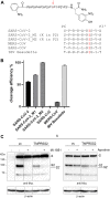
(A) Fluorescence resonance energy transfer substrates of the S protein S1/S2 sites of the indicated CoVs. M1 and M2 are mutants of the SARS-CoV-2 S1/S2 site with substitution of A → K or A → R in P2 position. IBV, avian infectious bronchitis virus strain Beaudette. Cleavage by furin is indicated in red. (B) Cleavage of the fluorescence resonance energy transfer substrates (20 μM) by furin (0.5 nM). Cleavage efficiency of SARS-CoV-2_M2 was set as 100%. (C) Cleavage of SARS-CoV-2 S by furin and TMPRSS2 in HEK293 cells. Cells were co-transfected with pCAGGS-S-Myc-6xHis and either empty vector or pCAGGS-TMPRSS2. Cells were then incubated in the absence or presence of aprotinin or furin inhibitor MI-1851 (50 μM each) for 48 h. Cell lysates were subjected to SDS–PAGE and Western blot analysis using antibodies against the C-terminal Myc-tag. For each Western blot lanes are spliced together from one immunoblot from one experiment. β-actin was used as loading control. Source data are available for this figure.
Figure S1.
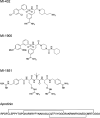
Figure S1.. Structural formulas of peptide mimetic inhibitors MI-432, MI-1900 and MI-1851 and the linear amino acid sequence of bovine aprotinin (24).
Aprotinin contains three disulfide bonds (indicated by lines).
Figure 3.
Figure 3.. Knockdown of TMPRSS2 expression by PPMO T-ex5 inhibits multicycle replication of SARS-CoV-2 in Calu-3 cells.
(A) Multicycle replication of SARS-CoV-2 in T-ex5–treated Calu-3 cells. Cells were treated with 25 μM T-ex5 or control PPMO (scramble) for 24 h or remained without treatment (w/o). Cells were then inoculated with SARS-CoV-2 at a MOI of 0.001 for 1 h 30 min, the inoculum was removed and the cells further incubated in the absence of PPMO for 72 h. Cells were fixed and immunostained using a serum against SARS-CoV. Virus-positive cells are stained in blue. Scale bars indicate 500 μm. (B) Calu-3 cells were treated with PPMO for 24 h and then infected with SARS-CoV-2 for 72 h as described above. Virus titers in supernatants were determined by tissue culture infection dose 50% (TCID50) end point dilution at indicated time points. Results are mean values ± SD of three independent experiments. (C) Analysis of TMPRSS2-mRNA in PPMO-treated Calu-3 cells. Cells were treated with 25 μM T-ex5, scramble PPMO or remained untreated (w/o) for 24 h (lanes 1–4). T-ex5–treated cells were inoculated with SARS-CoV-2 as described above and incubated in the absence of PPMO for 72 h (lane 4). Total RNA was isolated and analyzed by RT-PCR using primers designed to amplify 1,228 nt of full-length TMPRSS2-mRNA. Full-length and truncated PCR products lacking exon 5 are indicated by filled and open arrow heads, respectively. (D) Effect of PPMO treatment on Calu-3 cell viability. Calu-3 cells were treated with scramble or T-ex5 PPMO (25 μM) for 24 h. Cell viability of untreated (w/o) cells was set as 100%. Results are mean values ± SD (n = 3). Source data are available for this figure.
Figure 4.
Figure 4.. Inhibition of SARS-CoV-2 multiplication in human airway cells by inhibitors of furin and TMPRSS2.
Calu-3 cells were inoculated with SARS-CoV-2 at a low MOI of 0.001 and then incubated in the presence of inhibitors of TMPRSS2 (aprotinin, MI-432, and MI-1900), furin (MI-1851), and endosomal cathepsins (E64d), respectively, at the indicated concentrations. Cells were fixed and immunostained using a rabbit serum against SARS-CoV at 72 h p.i. Virus-positive cells are stained in blue or dark gray depending on the staining intensity. Cells infected in the absence of inhibitors (w/o), in the presence of DMSO (0.5%) and noninfected cells (mock) were used as controls. Scale bars indicate 500 μm. Images are representatives of three independent experiments.
Figure 5.
Figure 5.. Inhibition of SARS-CoV-2 multicycle replication in human airway epithelial cells by inhibitors of TMPRSS2 and furin.
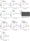
(A) Calu-3 cells were inoculated with SARS-CoV-2 at a low MOI of 0.001 and then incubated in the absence (w/o) or presence of inhibitors of TMPRSS2 (aprotinin, MI-432, and MI-1900), furin (MI-1851), and endosomal cathepsins (E64d), respectively, or DMSO (0.5%), at the indicated concentrations. At 16, 24, 48, and 72 h postinfection (p.i.), supernatants were collected, and virus replication was determined by tissue culture infection dose 50% (TCID50) titration at indicated time points. Data are mean values ± SD of three to five independent experiments. (B) Effect of inhibitor treatment on cell viability. Calu-3 cells were treated with the indicated protease inhibitor (50 μM) for 72 h. Untreated cells (w/o) and DMSO treated cells were used as controls. Cell viability of untreated cells was set as 100%. Results are mean values ± SD (n = 3). (C) Antiviral activity of combinations of TMPRSS2 and furin inhibitors against SARS-CoV-2 in human airway epithelial cells. Calu-3 cells were inoculated with SARS-CoV-2 at an MOI of 0.001 as described above and then incubated in the presence of single protease inhibitors or inhibitor combinations at the indicated concentrations. Virus titers in supernatants were determined by TCID50 at 16, 24, 48, and 72 h p.i. Data are mean values ± SD of three independent experiments. (D) Calu-3 cells were treated with PPMO for 24 h, then infected with SARS-CoV-2 as described above and incubated in the absence of PPMO (w/o, scramble and T-ex5) and with or without 50 μM of furin inhibitor treatment (MI-1851) for 72 h. At 16, 24, 48, and 72 h p.i., supernatants were collected, and viral titers were determined by TCID50 at indicated time points. Data are mean values ± SD (n = 2).
Figure S2.
Figure S2.. Cleavage analysis of SARS-CoV-2 S2′ site by furin.
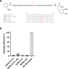
(A) Fluorescence resonance energy transfer substrates of the S protein S2′ sites of the indicated CoVs. M3 is a mutant of the SARS-CoV-2 S2′ site with substitution of P → R in P4 position. IBV, avian infectious bronchitis virus strain Beaudette. Cleavage by furin is indicated in red. (B) Cleavage of the fluorescence resonance energy transfer substrates (20 μM) by furin (0.5 nM). Cleavage efficiency of IBV Beaudette was set as 100%.
Figure 6.
Figure 6.. Proposed processing of SARS-CoV-2 spike protein S by TMPRSS2 and furin.
(i) S must be cleaved at two sites, S1/S2 and S2′, to trigger fusion of viral and cellular membranes during virus entry to release the virus genome into the host cell. CoV S cleavage is believed to occur sequentially, with cleavage at the S1/S2 site occurring first and subsequent cleavage at the S2′ site. Furin processes the S1/S2 site, whereas TMPRSS2 cleaves at the S2′ site, and both proteases cannot compensate each other. Inhibition of either furin (ii) or TMPRSS2 (iii) or simultaneous inhibition of both proteases (iv) renders the S protein fusion-inactive and prevents virus entry. Inhibition of TMPRSS2 prevents exposure of the fusion peptide at the N-terminus of the S2′ subunit (iii, iv). Inhibition of furin cleavage at the S1/S2 site may directly interfere with virus entry and membrane fusion by steric blockage of conformational changes (ii, upper scheme) or may prevent exposure of the S2′ site to TMPRSS2 (ii, lower scheme).

References

    1. Follis KK, York J, Nunberg JH (2006) Furin cleavage of the SARS coronavirus spike glycoprotein enhances cell-cell fusion but does not affect virion entry. Virology 350: 358–369. 10.1016/j.virol.2006.02.003 - DOI - PMC - PubMed
    1. Bosch BJ, Bartelink W, Rottier PJ (2008) Cathepsin L functionally cleaves the severe acute respiratory syndrome coronavirus class I fusion protein upstream of rather than adjacent to the fusion peptide. J Virol 82: 8887–8890. 10.1128/jvi.00415-08 - DOI - PMC - PubMed
    1. Belouzard S, Chu VC, Whittaker GR (2009) Activation of the SARS coronavirus spike protein via sequential proteolytic cleavage at two distinct sites. Proc Natl Acad Sci U S A 106: 5871–5876. 10.1073/pnas.0809524106 - DOI - PMC - PubMed
    1. Madu IG, Roth SL, Belouzard S, Whittaker GR (2009) Characterization of a highly conserved domain within the severe acute respiratory syndrome coronavirus spike protein S2 domain with characteristics of a viral fusion peptide. J Virol 83: 7411–7421. 10.1128/jvi.00079-09 - DOI - PMC - PubMed
    1. Walls AC, Tortorici MA, Bosch BJ, Frenz B, Rottier PJM, DiMaio F, Rey FA, Veesler D (2016) Cryo-electron microscopy structure of a coronavirus spike glycoprotein trimer. Nature 531: 114–117. 10.1038/nature16988 - DOI - PMC - PubMed

Publication types

Associated data

LinkOut - more resources