. 2020 Jul 24;369(6502):450-455.
doi: 10.1126/science.aaz1333.

Itaconate is an effector of a Rab GTPase cell-autonomous host defense pathway against Salmonella

Affiliations

Itaconate is an effector of a Rab GTPase cell-autonomous host defense pathway against Salmonella

Meixin Chen et al. Science. .

Abstract

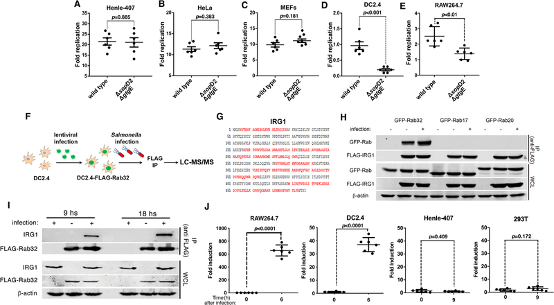
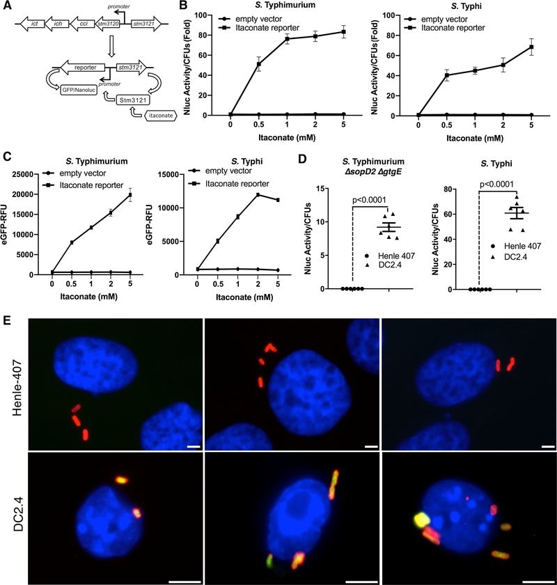
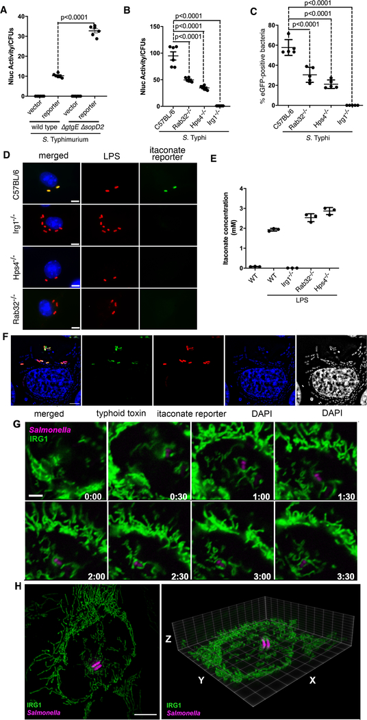
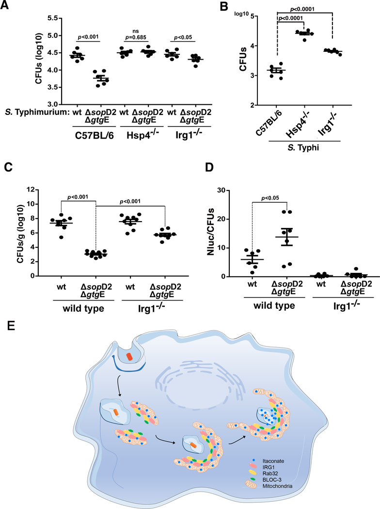
The guanosine triphosphatase (GTPase) Rab32 coordinates a cell-intrinsic host defense mechanism that restricts the replication of intravacuolar pathogens such as Salmonella Here, we show that this mechanism requires aconitate decarboxylase 1 (IRG1), which synthesizes itaconate, a metabolite with antimicrobial activity. We find that Rab32 interacts with IRG1 on Salmonella infection and facilitates the delivery of itaconate to the Salmonella-containing vacuole. Mice defective in IRG1 rescued the virulence defect of a S. enterica serovar Typhimurium mutant specifically defective in its ability to counter the Rab32 defense mechanism. These studies provide a link between a metabolite produced in the mitochondria after stimulation of innate immune receptors and a cell-autonomous defense mechanism that restricts the replication of an intracellular bacterial pathogen.


Conflict of interest statement

Competing interests: The authors declare no competing interests.

Figures

Fig. 1.
Fig. 1.. IRG1 interacts with Rab32 during Salmonella infection.
(A-E) The Rab32-associated pathogen restriction mechanism is manifested in myelocytic but not in epithelial cell lines. The ability of the S. Typhimurium ΔsopD2 ΔgtgE mutant strain to replicate within epithelial (Henle-407 and HeLa) or myelocytic (DC2.4 and RAW264.7) cell lines was evaluated by determining the CFU at different times after infection (MOI = 5). Fold replication represents the difference between the CFU at 1 and 9 hours post infection. Each circle represents the fold replication in each individual determination; the mean ± SEM of all the measurements and the p values of the indicated comparisons (two-sided Student’s t-test) are shown. (F-I) Rab32 interacts with IRG1 after Salmonella infection. DC2.4 cells expressing endogenous levels of FLAG-tagged Rab32 were infected with S. Typhimurium ΔsopD2 ΔgtgE (MOI = 30) and Rab32-interacting proteins were identified by affinity purification and LC—MS/MS analysis (F). The IRG1 peptides identified by the analyses are shown in red (G). (H and I) HEK293T cells transiently co-transfected with a plasmid expressing GFP-tagged Rab32, RAb17, or Rab20, along with a plasmid encoding FLAG-tagged IRG1 (H), or DC2.4 cells stably expressing FLAG-tagged Rab32 (I) were infected with S. Typhimurium ΔgtgE ΔsopD2 for 4 hours (MOI = 5). Cell lysates were then analyzed by immunoprecipitation with anti-FLAG and immunoblotting with anti-GFP, anti-FLAG, anti-IRG1, or anti-β-actin (as loading control) antibodies. IP: immunoprecipitates; WCL: whole-cell lysates. (J) Expression of IRG1 after Salmonella infection. The indicated cell lines were infected with S. Typhimurium ΔsopD2 ΔgtgE mutant strain (MOI = 5) and IRG1 mRNA levels were measured by qPCR 6 or 9 hours after infection. Each circle represents a single determination of the relative levels of IRG1 normalized to the levels of GAPDH; the mean ± SEM of all the measurements and p values of the indicated comparisons (two-sided Student’s t test) are shown.
Fig. 2.
Fig. 2.. Itaconate is delivered to the Salmonella-containing vacuole.
(A-C). Development of a biosensor to detect itaconate. (A) Chromosomal organization of the itaconate-degradation gene cluster in S. Typhimurium and diagram of the itaconate biosensor. (B and C) Effect of addition of itaconate on the biosensor transcriptional response. S. Typhimurium and S. Typhi strains carrying either the nanoluciferase or eGFP itaconate reporters were grown to an OD600 of 0.9 in the presence of different concentrations of itaconic acid (as indicated) and the levels of nanoluciferase or eGFP were determined. Values are the mean ± SD of three independent measurements. This experiment was repeated at least three times with equivalent results. (D and E) Detection of itaconate by intracellular Salmonella. DC2.4 or Henle-407 cells were infected with a S. Typhimurium ΔsopD2 ΔgtgE mutant (MOI=5) or S. Typhi (MOI=10) carrying a plasmid encoding a nanoluciferase-based itaconate biosensor. Eighteen hours after infection, the levels of nanoluciferase were measured in lysates of the infected cells (D). Each circle or triangle represents a single luciferase measurement; the mean ± SD and p values of the indicated comparisons (two-sided Student’s’s t test) are shown. This experiment was repeated at least three times with equivalent results. Alternatively, DC2.4 or Henle-407 cells were infected (MOI = 10) with S. Typhi carrying a plasmid encoding the eGFP-based itaconate biosensor (green). Eighteen hours after infection, cells were fixed, stained with DAPI (blue) to visualize nuclei, and stained with an anti-Salmonella LPS antibody along with Alexa 594-conjugated anti-rabbit antibody (red), and imaged under a fluorescence microscope (E). Scale bars: 5 μm.
Fig. 3.
Fig. 3.. Rab32/BLOC3-dependent delivery of itaconate to the Salmonella-containing vacuole.
(A) Cultured DC2.4 cells were infected with wild-type or ΔgtgE ΔsopD2 S. Typhimurium strains (MOI = 5) encoding the luciferase-based itaconate biosensor and the levels of luciferase in cell lysates were measured 9 hours after infection. Each circle represents a single luciferase measurement; the mean ± SD and the p-values of the indicated comparisons (two-sided Student’s t-test) are shown. (B-D) Bone-marrow-derived macrophages (BMDMs) obtained from C57BL/6, Rab32−/−, Hsp4−/−, or IRG1−/− mice were infected with S. Typhi (MOI = 10) encoding the luciferase- or eGFP-based itaconate biosensors. Nine hours after infection, the levels of luciferase in cell lysates (B) or the number of cells expressing eGFP (C) were determined. Each circle in (B) represents a single luciferase measurement. Values in (C) represent the percentage of bacterial cells exhibiting fluorescence. A minimum of 200 cells in each condition was evaluated. The mean ± SD and p-values of the indicated comparisons (one-way Anova) are shown. Representative fields of BMDMs obtained from the indicated mouse lines infected with S. Typhi encoding the eGFP itaconate reporter (green) are shown. Cells were fixed, stained with DAPI (blue) to visualize nuclei and stained with an anti-Salmonella LPS antibody along with Alexa 594-conjugated anti-rabbit antibody (red) (D) (scale bar = 5 μm). (E) Itaconate levels in BMDMs obtained from the indicated mice before and after LPS treatment to induce the expression of IRG1. Values represent the mean ± SD of three independent measurements. (F) Expression of the itaconate reporter (red) by intravacuolar but not by cytosolic S. Typhi. HeLa cells transfected with a plasmid encoding FLAG-tagged IRG1 were infected by a S. Typhi strain encoding a mCherry itaconate reporter (red) and a pltB::GFP transcriptional reporter (green). PltB, a component of S. Typhi’s typhoid toxin, is exclusively produced by bacteria located within the SCV and therefore serves as a surrogate to report for intravacuolar (GFP positive) vs intra cytosolic (GFP negative) bacteria. Six hours after infection, cells were stained with DAPI (to visualize all bacteria) and examined under a fluorescence microscope (scale bars: 5 μm). (G) Live-cell fluorescence time-lapse microscopy of cultured HeLa cells stably expressing IRG1-GFP (green) infected with S. Typhimurium ΔgtgE ΔsopD2 mutant strain encoding an mCherry itaconate biosensor (magenta). Imaging was initiated 45 minutes after infection. The times (hours:min) after initiation of imaging are indicated in each frame (the entire sequence is shown in video S1; this experiment was conducted at least three independent times, imaging several independent positions in each experiment, with equivalent findings; see videos S2 and S3 for additional examples). (H) Snapshot of a 3-D rendering of 3D-SIM acquisitions of HeLa cells stably expressing IRG1-GFP (green) infected with S. Typhimurium ΔgtgE ΔsopD2 mutant strain encoding an mCherry itaconate biosensor (magenta) (videos of this and additional reconstructions can be found in videos S4–S7).
Fig. 4.
Fig. 4.. Susceptibility of IRG1-deficient mice to Salmonella infection.
(A and B) Bone-marrow-derived macrophages (BMDMs) obtained from C57BL/6 (WT), Hsp4−/−, or IRG1−/− mice were infected with wild-type S. Typhimurium (MOI=5), its ΔgtgE ΔsopD2 mutant derivative (MOI=5) (A), or wild-type S. Typhi (MOI=10) (B), and the number of CFU was determined 9 hours after infection. Each circle represents the CFU in independent measurements; the mean ± SEM of all the measurements and p-values of the indicated comparisons (two-sided Student’s t test) are shown. (C and D) C57BL/6 (wild-type) or IRG1−/− mice were intraperitoneally infected with wild-type or ΔgtgE ΔsopD2 S. Typhimurium (as indicated) (102 CFU). Five days after infection, bacterial loads in the spleen of the infected animals were determined (C). Alternatively, mice were intraperitoneally infected with the same strains (104 CFU) and the levels of luciferase activity in spleen lysates was quantified 24 hours after infection (D). Each circle in (C) represents the bacterial loads of the spleen of an individual animal, and in (D) represents the luciferase levels in the spleen of an individual animal normalized to the CFU. The mean ± SEM of all the determination and p-values of the indicated comparisons (two-sided Student’s t-test) are shown. (E) Model for the mechanism of Rab32—BLOC3-mediated itaconate delivery to the Salmonella-containing vacuole. Upon infection, the mitochondrial network repositions to surround the incoming bacteria, and the resulting close interaction between the mitochondria and the Salmonella-containing vacuole results in the Rab32—BLOC3 dependent delivery of itaconate, which is synthesized in the mitochondria by IRG1.

References

    1. Randow F, MacMicking J, James L, Cellular self-defense: how cell-autonomous immunity protects against pathogens. Science 340, 701–706. (2013). - PMC - PubMed
    1. Reddick L, Alto N, Bacteria fighting back: how pathogens target and subvert the host innate immune system. Mol Cell. 54, 321–328 (2014). - PMC - PubMed
    1. Stenmark H, Rab GTPases as coordinators of vesicle traffic. Nat Rev Mol Cell Biol. 10, 513–525 (2009). - PubMed
    1. Spanò S, Galán J, A Rab32-dependent pathway contributes to Salmonella typhi host restriction. Science 338, 960–963 (2012). - PMC - PubMed
    1. Tang B, Rab32/38 and the xenophagic restriction of intracellular bacteria replication. Microbes Infect. 18, 595–603 (2016). - PubMed

Publication types