. 2020 Sep 18;369(6510):1505-1509.
doi: 10.1126/science.abc5881. Epub 2020 Jul 23.

Structural basis for neutralization of SARS-CoV-2 and SARS-CoV by a potent therapeutic antibody

Affiliations

Structural basis for neutralization of SARS-CoV-2 and SARS-CoV by a potent therapeutic antibody

Zhe Lv et al. Science. .

Abstract

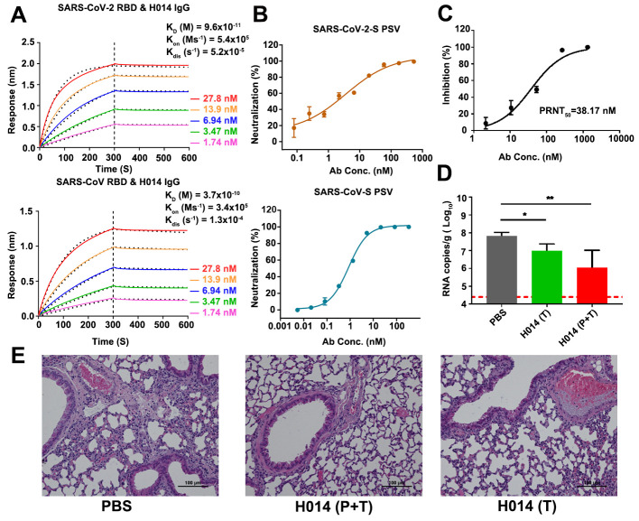
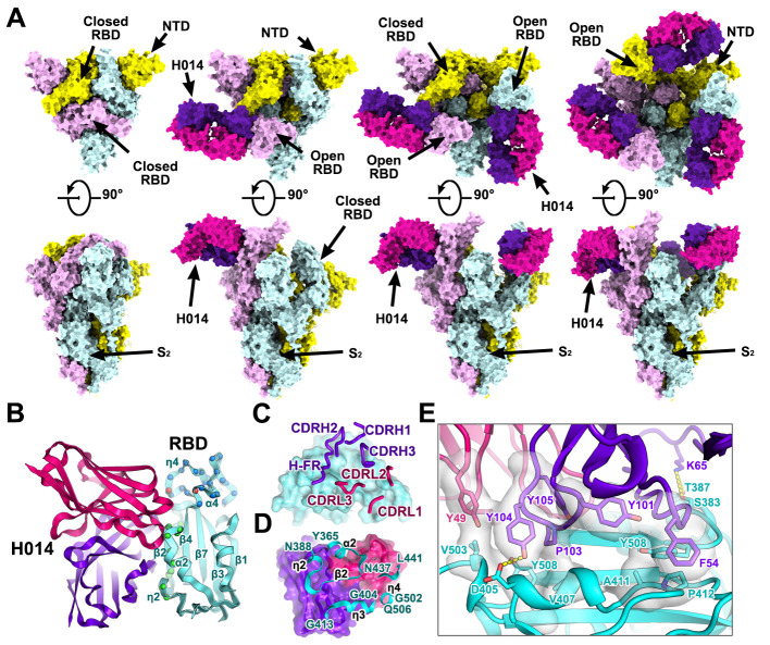
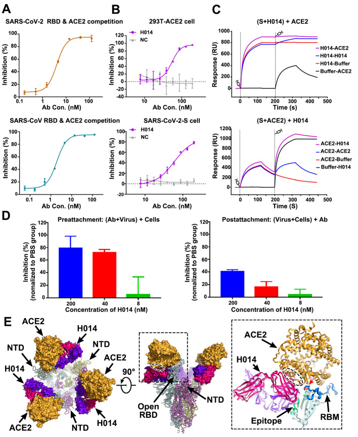
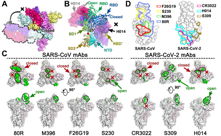
The coronavirus disease 2019 (COVID-19) pandemic caused by severe acute respiratory syndrome coronavirus 2 (SARS-CoV-2) has resulted in an unprecedented public health crisis. There are no approved vaccines or therapeutics for treating COVID-19. Here we report a humanized monoclonal antibody, H014, that efficiently neutralizes SARS-CoV-2 and SARS-CoV pseudoviruses as well as authentic SARS-CoV-2 at nanomolar concentrations by engaging the spike (S) receptor binding domain (RBD). H014 administration reduced SARS-CoV-2 titers in infected lungs and prevented pulmonary pathology in a human angiotensin-converting enzyme 2 mouse model. Cryo-electron microscopy characterization of the SARS-CoV-2 S trimer in complex with the H014 Fab fragment unveiled a previously uncharacterized conformational epitope, which was only accessible when the RBD was in an open conformation. Biochemical, cellular, virological, and structural studies demonstrated that H014 prevents attachment of SARS-CoV-2 to its host cell receptors. Epitope analysis of available neutralizing antibodies against SARS-CoV and SARS-CoV-2 uncovered broad cross-protective epitopes. Our results highlight a key role for antibody-based therapeutic interventions in the treatment of COVID-19.


Figures

Fig. 1
Fig. 1. H014 is a lineage B cross-neutralizing antibody of therapeutic value.
(A) Affinity analysis of the binding of H014 to RBD of SARS-CoV-2 and SARS-CoV. Biotinylated RBD proteins of (upper) SARS-CoV-2 or (lower) SARS-CoV were loaded on Octet SA sensor and tested for real-time association and dissociation of the H014 antibody. Global fit curves are shown as black dotted lines. The vertical dashed lines indicate the transition between association and disassociation phases. (B) Neutralizing activity of H014 against SARS-CoV-2 and SARS-CoV pseudoviruses (PSV). Serial dilutions of H014 were added to test its neutralizing activity against (upper) SARS-CoV-2 and (lower) SARS-CoV PSV. Neutralizing activities are represented as mean ± SD. Experiments were performed in triplicate. (C) In vitro neutralization activity of H014 against SARS-CoV-2 by PRNT in Vero cells. Neutralizing activities are represented as mean ± SD. Experiments were performed in duplicates. (D) Groups of hACE2 mice that received SARS-CoV-2 challenge were treated intraperitoneally with H014 in two independent experimental settings: 1) a single dose at 4 h post infection (Therapeutic, T); 2) two doses at 12 h before and 4 h post challenge (Prophylactic plus Therapeutic, P+T). Virus titers in the lungs were measured 5 days post infection (dpi) and are presented as RNA copies per gram of lung tissue. n=7/3/3, respectively. *P<0.05. LOD represents limit of detection. (E) Histopathological analysis of lung samples at 5 dpi. Scale bar: 100 μm.
Fig. 2
Fig. 2. Cryo-EM structures of the SARS-CoV-2 S trimer in complex with H014.
(A) Orthogonal views of SARS-CoV-2 S trimer with three RBDs in the closed state (left), one RBD in the open state and complexed with one H014 Fab (middle), two RBDs in the open state and each complexed with one H014 Fab. NTD: N-terminal domain. All structures are presented as molecular surfaces with different colors for each S monomer (cyan, violet and yellow), and the H014 Fab light (hotpink) and heavy (purpleblue) chains. (B) Cartoon representations of the structure of SARS-CoV-2 RBD in complex with H014 Fab with the same color scheme as in Fig. 2A. Residues comprising the H014 epitope and the RBM are shown as spheres and colored in green and blue, respectively. The overlapped residues between the H014 epitope and the RBM are shown in red. (C) and (D) Interactions between the H014 and SARS-CoV-2 RBD. The CDRs of the H014 that interact with SARS-CoV-2 RBD are displayed as thick tubes over the cyan surface of the RBD (C). The H014 epitope is shown as a cartoon representation over the surface of the RBD (D). (E) Details of the interactions between the H014 and SARS-CoV-2 RBD. Some residues involved in the formation of hydrophobic patches and hydrogen bonds are shown as sticks and labeled. Color scheme is the same as in Fig. 2A.
Fig. 3
Fig. 3. Mechanism of neutralization of H014.
(A) Competitive binding assays by ELISA. Recombinant SARS-CoV-2 (upper) or SARS-CoV (lower) RBD protein was coated on 96-well plates, recombinant ACE2 and serial dilutions of H014 were then added for competitive binding to SARS-CoV-2 or SARS-CoV RBD. Values are mean ± SD. Experiments were performed in triplicate. (B) Blocking of SARS-CoV-2 RBD binding to 293T-ACE2 cells by H014 (upper). Recombinant SARS-CoV-2 RBD protein and serially diluted H014 were incubated with ACE2 expressing 293T cells (293T-ACE2) and tested for binding of H014 to 293T-ACE2 cells. Competitive binding of H014 and ACE2 to SARS-CoV-2-S cells (lower). Recombinant ACE2 and serially diluted H014 were incubated with 293T cells expressing SARS-CoV-2 Spike protein (SARS-CoV-2-S) and tested for binding of H014 to SARS-CoV-2-S cells. BSA was used as a negative control (NC). Values are mean ± SD. Experiments were performed in triplicate. (C) BIAcore SPR kinetics of competitive binding of H014 and ACE2 to SARS-CoV-2 S trimer. For both panels, SARS-CoV-2 S trimer was loaded onto the sensor. In the upper panel, H014 was first injected, followed by ACE2, whereas in the lower panel, ACE2 was injected first and then H014. The control groups are depicted by black curves. (D) Amount of virus on the cell surface, as detected by RT-PCR. Pre-attachment mode: incubate SARS-CoV-2 and H014 first, then add the mixture into cells (left); post-attachment mode: incubate SARS-CoV-2 and cells first, then add H014 into virus-cell mixtures (right). High concentrations of H014 prevent attachment of SARS-CoV-2 to the cell surface when SARS-CoV-2 was exposed to H014 before cell attachment. Values represent mean ± SD. Experiments were performed in duplicates. (E) Clashes between H014 Fab and ACE2 upon binding to SARS-CoV-2 S. H014 and ACE2 are represented as surface; SARS-CoV-2 S trimer is shown as ribbon. Inset is a zoomed-in view of the interactions of the RBD, H014 and ACE2 and the clashed region (oval ellipse) between H014 and ACE2. The H014 Fab light and heavy chains, ACE2, and RBD are presented as cartoons. The epitope, RBM and the overlapped binding region of ACE2 and H014 on RBD are highlighted in green, blue and red, respectively.
Fig. 4
Fig. 4. Breathing of the S1 subunit and epitopes of neutralizing antibodies.
(A) H014 can only interact with the “open” RBD, whereas the “closed” RBD is inaccessible to H014. The “open” RBD and RBD bound H014 are depicted in lighter colors corresponding to the protein chain they belong to. Color scheme is the same as in Fig. 2A. (B) Structural rearrangements of the S1 subunit of SARS-CoV-2 transition from the closed state to the open state. SD1: subdomain 1, SD2: subdomain 2, RBD’: RBD (closed state) from adjacent monomer. SD1, SD2, NTD, RBD and RBD’ are colored in pale green, light orange, cyan, blue and yellow, respectively. The red dot indicates the hinge point. The angles between the RBD and SD1 are labeled. (C) Epitope location analysis of neutralizing antibodies on SARS-CoV and SARS-CoV-2 S trimers. The S trimer structures with one RBD open and two RBD closed from SARS-CoV and SARS-CoV-2 were used to show individual epitope information, which is highlighted in green. The accessible and in-accessible states are encircled and marked by green ticks and red crosses. (D) Footprints of the seven mAbs on RBDs of SARS-CoV (left) and SARS-CoV-2 (right). RBDs are rendered as molecular surfaces in light blue. Footprints of different mAbs are highlighted in different colors as labeled in the graph. Note: epitopes recognized by indicated antibodies are labeled in blue (non-overlapped), yellow (overlapped once) and red (overlapped twice).

References

    1. Zhou P., Yang X.-L., Wang X.-G., Hu B., Zhang L., Zhang W., Si H.-R., Zhu Y., Li B., Huang C.-L., Chen H.-D., Chen J., Luo Y., Guo H., Jiang R.-D., Liu M.-Q., Chen Y., Shen X.-R., Wang X., Zheng X.-S., Zhao K., Chen Q.-J., Deng F., Liu L.-L., Yan B., Zhan F.-X., Wang Y.-Y., Xiao G.-F., Shi Z.-L., A pneumonia outbreak associated with a new coronavirus of probable bat origin. Nature 579, 270–273 (2020). 10.1038/s41586-020-2012-7 - DOI - PMC - PubMed
    1. Walls A. C., Park Y.-J., Tortorici M. A., Wall A., McGuire A. T., Veesler D., Structure, Function, and Antigenicity of the SARS-CoV-2 Spike Glycoprotein. Cell 181, 281–292.e6 (2020). 10.1016/j.cell.2020.02.058 - DOI - PMC - PubMed
    1. Gao Q., Bao L., Mao H., Wang L., Xu K., Yang M., Li Y., Zhu L., Wang N., Lv Z., Gao H., Ge X., Kan B., Hu Y., Liu J., Cai F., Jiang D., Yin Y., Qin C., Li J., Gong X., Lou X., Shi W., Wu D., Zhang H., Zhu L., Deng W., Li Y., Lu J., Li C., Wang X., Yin W., Zhang Y., Qin C., Development of an inactivated vaccine candidate for SARS-CoV-2. Science 369, 77–81 (2020). 10.1126/science.abc1932 - DOI - PMC - PubMed
    1. Corti D., Zhao J., Pedotti M., Simonelli L., Agnihothram S., Fett C., Fernandez-Rodriguez B., Foglierini M., Agatic G., Vanzetta F., Gopal R., Langrish C. J., Barrett N. A., Sallusto F., Baric R. S., Varani L., Zambon M., Perlman S., Lanzavecchia A., Prophylactic and postexposure efficacy of a potent human monoclonal antibody against MERS coronavirus. Proc. Natl. Acad. Sci. U.S.A. 112, 10473–10478 (2015). 10.1073/pnas.1510199112 - DOI - PMC - PubMed
    1. Pallesen J., Wang N., Corbett K. S., Wrapp D., Kirchdoerfer R. N., Turner H. L., Cottrell C. A., Becker M. M., Wang L., Shi W., Kong W.-P., Andres E. L., Kettenbach A. N., Denison M. R., Chappell J. D., Graham B. S., Ward A. B., McLellan J. S., Immunogenicity and structures of a rationally designed prefusion MERS-CoV spike antigen. Proc. Natl. Acad. Sci. U.S.A. 114, E7348–E7357 (2017). 10.1073/pnas.1707304114 - DOI - PMC - PubMed

Publication types

MeSH terms