Review
. 2020 Sep;16(9):489-508.
doi: 10.1038/s41581-020-0309-2. Epub 2020 Jul 23.

Autophagy in kidney homeostasis and disease

Affiliations
Review

Autophagy in kidney homeostasis and disease

Chengyuan Tang et al. Nat Rev Nephrol. 2020 Sep.

Abstract

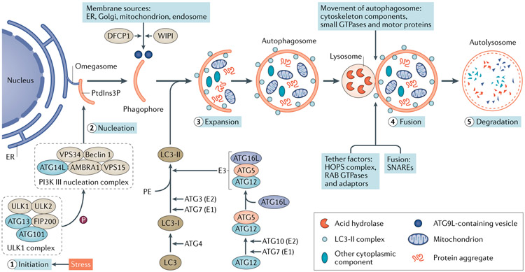
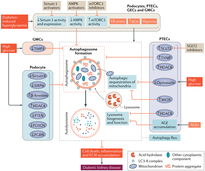
Autophagy is a conserved lysosomal pathway for the degradation of cytoplasmic components. Basal autophagy in kidney cells is essential for the maintenance of kidney homeostasis, structure and function. Under stress conditions, autophagy is altered as part of the adaptive response of kidney cells, in a process that is tightly regulated by signalling pathways that can modulate the cellular autophagic flux - mammalian target of rapamycin, AMP-activated protein kinase and sirtuins are key regulators of autophagy. Dysregulated autophagy contributes to the pathogenesis of acute kidney injury, to incomplete kidney repair after acute kidney injury and to chronic kidney disease of varied aetiologies, including diabetic kidney disease, focal segmental glomerulosclerosis and polycystic kidney disease. Autophagy also has a role in kidney ageing. However, questions remain about whether autophagy has a protective or a pathological role in kidney fibrosis, and about the precise mechanisms and signalling pathways underlying the autophagy response in different types of kidney cells and across the spectrum of kidney diseases. Further research is needed to gain insights into the regulation of autophagy in the kidneys and to enable the discovery of pathway-specific and kidney-selective therapies for kidney diseases and anti-ageing strategies.


Figures

Fig. 1 ∣
Fig. 1 ∣. Autophagy dynamics and core machinery.
Autophagy is a muitistep process involving initiation, nucleation, expansion, fusion and degradation. Upon induction, the ULK1 complex — comprising the serine/threonine protein kinases ULK1 and ULK2, autophagy-related protein 13 (ATG13), FAK family kinase-interacting protein of 200 kDa (FIP200) and ATG101 — phosphorylates multiple substrates, including components of the phosphoinositide 3-kinase (PI3K) III nucleation complex to promote phagophore nucleation. The PI3K III complex comprises PI3K catalytic subunit type 3 (VPS34), PI3K regulatory subunit 4 (VPS15), beclin 1, beclin 1-associated autophagy-related key regulator (ATG14L) and activating molecule in beclin 1-regulated autophagy protein 1 (AMBRA1). This complex produces phosphatidylinositol-3-phosphate (PtdIns3P) at the omegasome, which recruits PtdIns3P-binding proteins for phagophore expansion. The delivery of membrane from other sources to the expanding phagophore requires PtdIns3P-binding proteins WD repeat domain phosphoinositide-interacting proteins (WIPIs) and zinc-finger FYVE domain-containing protein 1 (DFCP1), as well as cycling of the transmembrane protein ATG9L in the form of membrane vesicles and tubules. Two ubiquitin-like conjugation systems, the ATG12–ATG5–ATG16L system and the microtubule-associated protein 1 light chain 3 (LC3) system, participate in autophagosome elongation and completion. The conversion of cytosolic LC3-I into membrane-bound LC3-II is indicative of autophagy induction and autophagosome formation. The completed autophagosome then fuses with a lysosome to form an autolysosome, in which the autophagosome inner membrane and cargos are degraded by lysosomal hydrolases and eventually released for recycling. The fusion process is regulated by a large set of molecules, including cytoskeleton components and related motor proteins, tethering factors including the homotypic fusion and vacuole protein sorting (HOPS) complex, the RAB GTPases, and specific soluble N-ethylmaleimide-sensitive factor attachment protein receptor (SNARE) complexes. E1, ubiquitin-activating enzyme; E2, ubiquitin-conjugating enzyme; E3, ubiquitin ligase; ER, endoplasmic reticulum; PE, phosphatidylethanolamine.
Fig. 2 ∣
Fig. 2 ∣. Mitophagy pathways and cargo recognition.
Mitophagy is mainly mediated by microtubule-associated protein 1 light chain 3 (LC3)-associated autophagy receptors via both ubiquitin (Ub)-dependent and Ub-independent pathways. Ub-dependent mitophagy involves the mitochondrial serine/threonine protein kinase PINK1 and E3 Ub-protein ligase parkin (PINK1–parkin) pathway. Following loss of mitochondrial membrane potential, PINK1 accumulates on the outer mitochondrial membrane (OMM), where it recruits and phosphorylates parkin to add phospho–Ub chains on OMM proteins. Autophagy receptors, such as sequestosome 1, next to BRCA1 protein (NBR1), optineurin, calcium-binding and coiled-coil domain-containing protein 2 (NDP52) and TAX1-binding protein 1 (TAX1BP1), which contain both Ub-binding domains and LC3-interacting regions (LIRs), bridge the ubiquitylated mitochondria to LC3-associated autophagosomal membranes for sequestration. PINK1-mediated phosphorylation of ubiquitin might be sufficient to recruit NDP52 and optineurin and induce mitophagy independently of parkin. Ub-independent mitophagy is mediated by several OMM receptors such as BCL-2/adenovirus E1B 19 kDa protein-interacting protein 3 (BNIP3), BNIP3-like (BNIP3L), FUN14 domain-containing protein 1 (FUNDC1), which do not require interaction with Ub for cargo recognition. Peptidyl-prolyl cis-trans isomerase FKBP8 and BCL-2-like 13 (BCL2L13) are potential additional mitophagy receptors. By binding to LC3 via the LIRs at their cytosolic N terminus, mitophagy receptors link damaged mitochondria directly to autophagosomes.
Fig. 3 ∣
Fig. 3 ∣. Signalling networks for the induction of autophagy.
Mammalian target of rapamycin complex 1 (mTORC1) is the major negative regulator of autophagy, whereas AMP-activated protein kinase (AMPK) and sirtuin 1 are positive regulators. mTOR is activated by amino acids via the RAG family of GTPases or by growth factors through two protein kinase pathways that involve RACα serine/threonine protein kinase (AKT) and mitogen-activated protein kinase kinase (MAPKK)–extracellular signal-regulated kinase (ERK). AKT and ERK1 or ERK2 can phosphorylate the hamartin–tuberin complex, which negatively regulates mTORC1 by inhibiting GTP-binding protein RHEB. AMPK is phosphorylated and activated by several upstream kinases, including serine/threonine protein kinase STK11, calcium/calmodulin-dependent protein kinase 2 (CAMKK2) and mitogen-activated protein kinase kinase kinase 7 (TAK1). Once activated, AMPK can directly phosphorylate serine/threonine protein kinase ULK1 to promote autophagy and/or phosphorylate hamartin–tuberin and regulatory-associated protein of mTOR (RAPTOR), which is one of the components of mTORC1, to inhibit mTORC1 and enable the induction of autophagy. Depletion of cellular energy stores increases NAD+ levels and activates sirtuin 1. Active sirtuin 1 induces autophagy by directly deacetylating several essential autophagy proteins such as autophagy-related protein 5 (ATG5), ATG7 and microtubule-associated protein 1 light chain 3 (LC3). During starvation, sirtuin 1 can deacetylate forkhead box protein O1 (FOXO1) to upregulate the GTPase RAB7 and promote autophagic flux. Sirtuin 1 can also deacetylate FOXO3 to enhance the expression of various genes that encode ATGs such as ULK2, beclin 1, phosphoinositide 3-kinase catalytic subunit type 3 (also known as VPS34), BCL-2/adenovirus E1B 19 kDa protein-interacting protein 3 (BNIP3), BNIP3-like (BNIP3L), ATG12, ATG4B and LC3. Moreover, sirtuin 1 promotes autophagy by interacting with hamartin–tuberin and deacetylating STK11. mTOR-independent pathways have also been implicated in autophagic regulation under conditions of nutrient shortage. Upon starvation, mitogen-activated protein kinase 8 (MAPK8) is activated, which phosphorylates the apoptosis regulator BCL-2 to liberate and activate beclin 1 to induce autophagy. Activated MAPK8 might also phosphorylate sirtuin 1 to promote its enzymatic activity. Phosphorylation of beclin 1 by death-associated protein kinases (DAPKs) facilitates its dissociation from the anti-apoptotic protein BCL2L1, which also promotes the induction of autophagy. Activation of eukaryotic translation initiation factor 2 subunit-α (eIF2α) and of inhibitor of NF-κB kinase (IKK) is also involved in starvation-induced autophagy. In addition, starvation stimulates the nuclear translocation of transcription factor EB (TFEB) to transactivate the expression of genes involved in autophagosome formation, lysosome biogenesis and lysosome function. By contrast, starvation stimulates the nuclear export of zinc-finger protein with KRAB and SCAN domains 3 (ZKSCAN3), which prevents its repression of ATG gene expression. ATF4, activating transcription factor 4; PI3K, phosphoinositide 3-kinase; RAF, RAF proto-oncogene serine/threonine-protein kinase.
Fig. 4 ∣
Fig. 4 ∣. Autophagy in AKI and kidney interstitial fibrosis.
Acute kidney injury (AKI) induces autophagy in proximal tubule epithelial cells (PTECs) to protect against kidney injury. Following the initial injury, autophagy in PTECs undergoes dynamic changes and mammalian target of rapamycin (mTOR)-mediated autophagy resolution during the injury recovery phase facilitates tubular cell proliferation and repair. Moreover, tightly regulated autophagy is required for phagocytosis of apoptotic cells by epithelial cells, which might also inhibit inflammatory responses and thus promote kidney repair. By contrast, persistent induction of autophagy in PTECs after AKI might induce cellular senescence and suppress cell proliferation. These autophagy-mediated changes stimulate the production and secretion of pro-fibrotic cytokines, leading to maladaptive repair and tubulointerstitial fibrosis. mTORC, mTOR complex.
Fig. 5 ∣
Fig. 5 ∣. Autophagy in diabetic kidney disease.
Diabetes-associated hyperglycaemia induces cellular stress, including an increase in reactive oxygen species (ROS), endoplasmic reticulum (ER) stress and hypoxia. In the kidney, the initial adaptive response seems to include the activation of autophagy in podocytes, proximal tubule epithelial cells (PTECs), glomerular endothelial cells (GECs) and glomerular mesangial cells (GMCs). By contrast, sustained disturbance of major nutrient and/or energy sensing pathways leads to activation of mammalian target of rapamycin complex 1 (mTORC1), and inhibition of AMP-activated protein kinase (AMPK) and sirtuin 1, which suppresses autophagy in these kidney cells. In podocytes exposed to high glucose, increased expression of β-arrestins or histone deacetylase 4 (HDAC4), or reduced expression of phosphatase and tensin homologue (PTEN), forkhead box protein O1 (FOXO1), progranulin (PGRN), sirtuin 6 or tyrosine-protein phosphatase non-receptor type substrate 1 (SIRPα) might result in autophagy impairment. In PTECs, exposure to high glucose increased the expression of sodium–glucose cotransporter 2 (SGLT2), thioredoxin-interacting protein (TXNIP), HDAC6 and inositol oxygenase (MIOX), or reduced expression of optineurin, all of which might also impair autophagy. Moreover, accumulation of advanced glycation end-products (AGEs) in PTECs might cause lysosome dysfunction and consequently disrupt auto-lysosome formation and function. In GMCs, reduced expression of metalloproteinase inhibitor 3 (TIMP3) might inhibit autophagy. Collectively, intracellular stress, disturbances of nutrient-sensing pathways and changes in these signalling pathways might lead to dysregulated autophagy and the progression of DKD. ECM, extracellular matrix; LC3, microtubule-associated protein 1 light chain 3.

References

    1. Dikic I & Elazar Z Mechanism and medical implications of mammalian autophagy. Nat. Rev. Mol. Cell Biol 19, 349–364 (2018). - PubMed
    1. Oku M & Sakai Y Three distinct types of microautophagy based on membrane dynamics and molecular machineries. Bioessays 40, e1800008 (2018). - PubMed
    1. Kaushik S & Cuervo AM The coming of age of chaperone-mediated autophagy. Nat. Rev. Mol. Cell Biol 19, 365–381 (2018). - PMC - PubMed
    1. Levine B & Kroemer G Biological functions of autophagy genes: a disease perspective. Cell 176, 11–42 (2019). - PMC - PubMed
    1. Morishita H & Mizushima N Diverse cellular roles of autophagy. Annu. Rev. Cell Dev. Biol 35, 453–475 (2019). - PubMed

Publication types

MeSH terms