. 2021 Jan;28(1):203-218.
doi: 10.1038/s41418-020-0592-2. Epub 2020 Jul 23.

Intranasal delivery of mesenchymal stem cell secretome repairs the brain of Alzheimer's mice

Affiliations

Intranasal delivery of mesenchymal stem cell secretome repairs the brain of Alzheimer's mice

Giulia Santamaria et al. Cell Death Differ. 2021 Jan.

Abstract

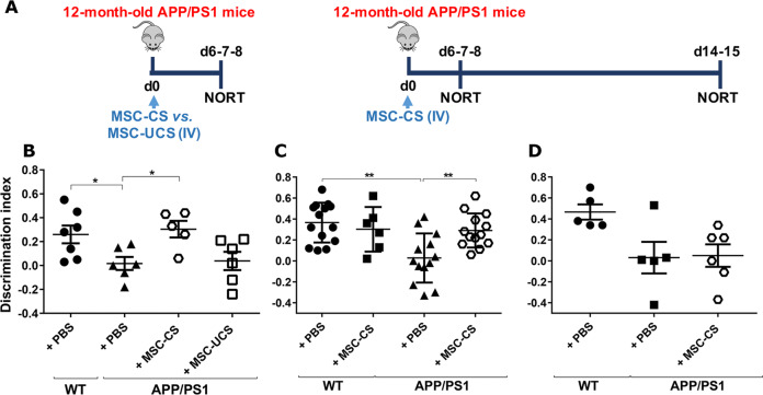
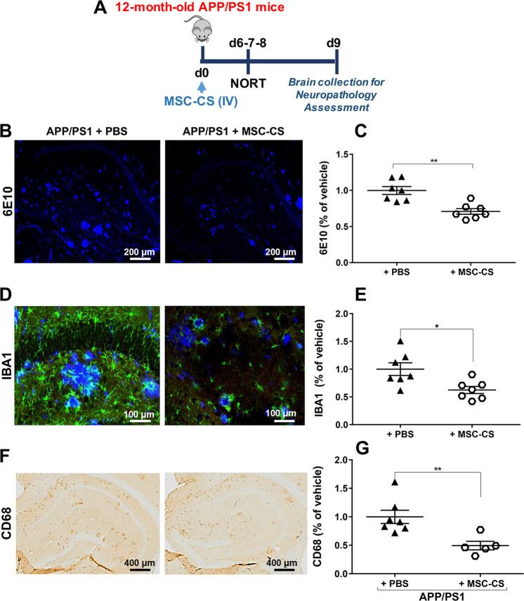
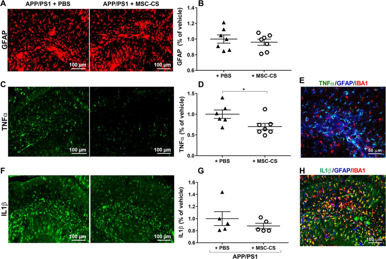
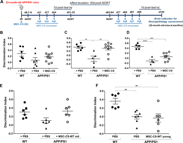
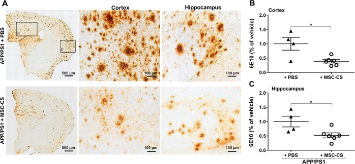
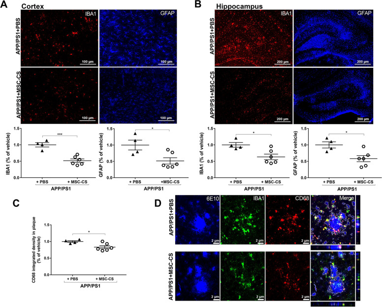
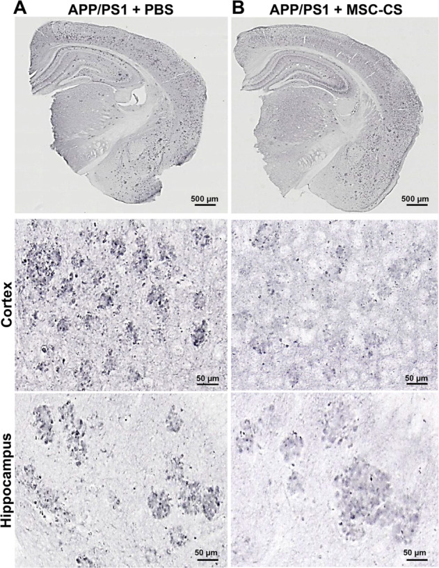
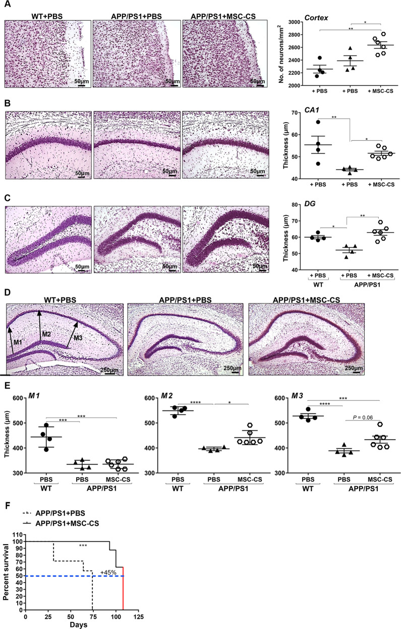
The multiplicity of systems affected in Alzheimer's disease (AD) brains calls for multi-target therapies. Although mesenchymal stem cells (MSC) are promising candidates, their clinical application is limited because of risks related to their direct implantation in the host. This could be overcome by exploiting their paracrine action. We herein demonstrate that in vivo systemic administration of secretome collected from MSC exposed in vitro to AD mouse brain homogenates (MSC-CS), fully replicates the cell-mediated neuroreparative effects in APP/PS1 AD mice. We found a complete but transient memory recovery by 7 days, which vanished by 14 days, after a single MSC-CS intravenous administration in 12-month or 22-24-month-old mice. Treatment significantly reduced plaque load, microglia activation, and expression of cytokines in astrocytes in younger, but not aged, mice at 7 days. To optimize efficacy, we established a sustained treatment protocol in aged mice through intranasal route. Once-weekly intranasal administration of MSC-CS induced persistent memory recovery, with dramatic reduction of plaques surrounded by a lower density of β-amyloid oligomers. Gliosis and the phagocytic marker CD68 were decreased. We found a higher neuronal density in cortex and hippocampus, associated with a reduction in hippocampal shrinkage and a longer lifespan indicating healthier conditions of MSC-CS-treated compared to vehicle-treated APP/PS1 mice. Our data prove that MSC-CS displays a great multi-level therapeutic potential, and lay the foundation for identifying the therapeutic secretome bioreactors leading to the development of an efficacious multi-reparative cocktail drug, towards abrogating the need for MSC implantation and risks related to their direct use.


Conflict of interest statement

The authors declare that they have no conflict of interest.

Figures

Fig. 1
Fig. 1. One IV injection of MSC-CS, but not MSC-UCS, transiently restores memory in 12-month-old APP/PS1 mice.
a Experimental design. b Comparison of the discrimination index (DI) of APP/PS1 mice and their age-matched WT littermates receiving either PBS, MSC-CS or MSC-UCS and tested in the NORT. DI of WT and APP/PS1 mice treated with PBS or MSC-CS and tested in the NORT 7 days (c) and 14 days (d) post-injection. Data are expressed as scatter plots with mean ± SEM. One-way ANOVA in b and d. Two-way ANOVA in c, *P < 0.05; **P < 0.01 Tukey’s multiple comparison post-hoc test.
Fig. 2
Fig. 2. A single IV injection of MSC-CS reduces brain amyloidosis and microglial activation.
a Experimental design. b Confocal images of the hippocampal area marked with the 6E10 anti-Aβ antibody, evidencing deposited amyloid plaques. c Plaque quantification. d Confocal images of the hippocampal area marked with both 6E10 for plaques and IBA1 for microglial cells. e IBA1-marked area quantification. f Hippocampal area immunostained for the phagocytic marker CD68 quantified in g. Data are expressed as scatter plots with mean ± SEM. *P < 0.05; **P < 0.01 Student’s t test.
Fig. 3
Fig. 3. Single IV injection of MSC-CS improves the inflammatory phenotype of astrocytes.
a Confocal images of hippocampal slices from MSC-CS-treated and PBS-treated mice immunostained for GFAP. b GFAP-marked area quantification evidencing the degree of astrogliosis between treatment groups. c Hippocampal slices immunostained for TNFα and relative quantification in d. e Merge of hippocampal immunostaining for TNFα-GFAP-IBA1. f Hippocampal immunostaining for IL1β quantified in g; h Merge of hippocampal immunostaining for IL1β-GFAP-IBA1. Quantification data are expressed as scatter plots with mean ± SEM. *P < 0.05, Student’s t test.
Fig. 4
Fig. 4. Repeated IN delivery of MSC-CS significantly restores memory whereas MSC-CS-WT old or young has partial or no effect, respectively.
a Experimental design for MSC-CS treatment. DI of the recognition memory task of mice tested 7 days after 1 (b) or 4 (c) IN administrations, or 39 days after suspension of MSC-CS treatment (d). e DI of the recognition memory task of mice tested 7 days after 4 IN administrations of MSC-CS-WT old. f DI of the recognition memory task of mice tested 7 days after 4 IN administrations of MSC-CS-WT young. Data are expressed as scatter plots with mean ± SEM. One-way ANOVA; **P < 0.01; ***P < 0.001; ****P < 0.0001, Tukey’s multiple comparison post-hoc test.
Fig. 5
Fig. 5. Repeated IN delivery of MSC-CS significantly decreases brain amyloidosis.
a 6E10 immunostaining comparing brain plaque load between MSC-CS- and PBS-treated mice (left panels). In the middle and right panels, higher magnification of plaques in cortex (c) and hippocampus (h) showing MSC-CS-mediated changes in terms of aggregate density, and absence of the central plaque core. b, c Plaque quantification in the cortex and the hippocampus. Quantitative data are expressed as scatter plots with mean ± SEM. *P < 0.05, Student’s t test.
Fig. 6
Fig. 6. Repeated IN injections of MSC-CS significantly reduce neuroinflammation.
a, b IBA1 and GFAP immunostaining showing the extent of gliosis in the cortex and the hippocampus between PBS- and MSC-CS-treated (8 IN) 25-month-old APP/PS1 mice (upper panels). Quantification of the IBA1- and GFAP-marked areas in the cortex and the hippocampus, respectively (lower panels). c CD68 quantification in plaques. d Immunofluorescence images of plaques (6E10), CD68 and IBA1 expression, and CD68-IBA1 co-localization (Merge) around plaques between treatment groups. Quantitative data are expressed as scatter plots with mean ± SEM. ***P < 0.001; *P < 0.05, Student’s t test.
Fig. 7
Fig. 7. Repeated delivery of MSC-CS appears to reduce AβO load around plaques in APP/PS1 mice.
Images of APP/PS1 mouse brain slices immunostained with the anti-AβOs A11 antibody showing the presence of AβOs in PBS- (a) and MSC-CS-treated (8 IN) 25-month-old mice (b); upper panels. Middle and lower panels depict a higher magnification of plaques in the cortex and the hippocampus of APP/PS1 mice showing AβO distribution (darker dots).
Fig. 8
Fig. 8. APP/PS1 mice treated repeatedly with MSC-CS display a higher number of neuronal cells in the cortex and the hippocampus, reduced hippocampal atrophy, and increased survival.
Left panels are representative NISSL-stained sections of the cortex (a) and cell layer thickness in the CA1 region of the hippocampus (b) and the dentate gyrus (DG) (c). Right panels show neuronal quantification thereof. d Images of NISSL-stained brain slices in which headed arrows at M1, M2 and M3 indicate where thickness was measured between the CA1 and DG hippocampal sub-regions. e Quantification of neuropil thickness for the 3 zones selected. Data are expressed as scatter plots with mean ± SEM. One-way ANOVA; *P < 0.05, **P < 0.01; ***P < 0.001, ****P < 0.0001; Tukey’s multiple comparison post-hoc test. f The graph describes mouse longevity comparison between MSC-CS-treated and PBS-treated APP/PS1 mice. The red line in APP/PS1 + MSC-CS indicates that these mice were sacrificed for experimental needs, but were still perfectly healthy. ***P < 0.001, Student’s t test.

References

    1. Selkoe DJ, Hardy J. The amyloid hypothesis of Alzheimer’s disease at 25 years. EMBO Mol Med. 2016;8:595–608. doi: 10.15252/emmm.201606210. - DOI - PMC - PubMed
    1. Selkoe DJ. Alzheimer’s disease. Cold Spring Harb Perspect Biol. 2011;3:a004457. doi: 10.1101/cshperspect.a004457. - DOI - PMC - PubMed
    1. Selkoe DJ. Alzheimer’s disease is a synaptic failure. Science. 2002;298:789–91. doi: 10.1126/science.1074069. - DOI - PubMed
    1. Benilova I, Karran E, De Strooper B. The toxic Abeta oligomer and Alzheimer’s disease: an emperor in need of clothes. Nat Neurosci. 2012;15:349–57. doi: 10.1038/nn.3028. - DOI - PubMed
    1. Heneka MT, Carson MJ, El Khoury J, Landreth GE, Brosseron F, Feinstein DL, et al. Neuroinflammation in Alzheimer’s disease. Lancet Neurol. 2015;14:388–405. doi: 10.1016/S1474-4422(15)70016-5. - DOI - PMC - PubMed

Publication types