. 2020 Aug 1;143(8):2576-2593.
doi: 10.1093/brain/awaa179.

Impaired glymphatic function and clearance of tau in an Alzheimer's disease model

Affiliations

Impaired glymphatic function and clearance of tau in an Alzheimer's disease model

Ian F Harrison et al. Brain. .

Abstract

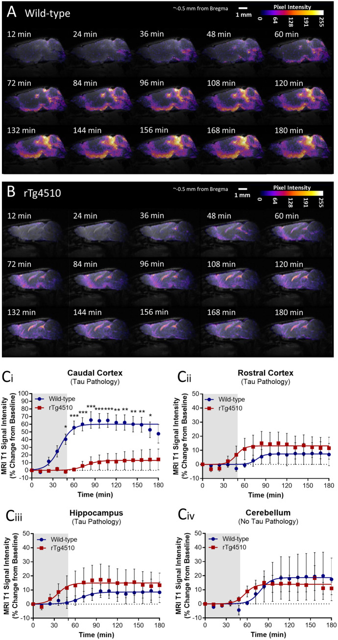
The glymphatic system, that is aquaporin 4 (AQP4) facilitated exchange of CSF with interstitial fluid (ISF), may provide a clearance pathway for protein species such as amyloid-β and tau, which accumulate in the brain in Alzheimer's disease. Further, tau protein transference via the extracellular space, the compartment that is cleared by the glymphatic pathway, allows for its neuron-to-neuron propagation, and the regional progression of tauopathy in the disorder. The glymphatic system therefore represents an exciting new target for Alzheimer's disease. Here we aim to understand the involvement of glymphatic CSF-ISF exchange in tau pathology. First, we demonstrate impaired CSF-ISF exchange and AQP4 polarization in a mouse model of tauopathy, suggesting that this clearance pathway may have the potential to exacerbate or even induce pathogenic accumulation of tau. Subsequently, we establish the central role of AQP4 in the glymphatic clearance of tau from the brain; showing marked impaired glymphatic CSF-ISF exchange and tau protein clearance using the novel AQP4 inhibitor, TGN-020. As such, we show that this system presents as a novel druggable target for the treatment of Alzheimer's disease, and possibly other neurodegenerative diseases alike.

Keywords: Alzheimer’s disease; aquaporin-4; glymphatic; rTg4510; tau.


Figures

Figure 1
Figure 1
Heterogeneous deposition of tau in the cortex of rTg4510 mice. (A) NeuN and (B) CaMK2a expression in the mouse brain appear homogenous throughout rostral to caudal aspects of the cortex. During early deposition of tau in the rTg4510 mouse brain, however, pTau (PG-5) staining appears greater in the rostral compared to the caudal cortex, at (C) 3.2 months and (D) 3.7 months, subsiding by (E) 4.6 months of age. Subiculum of the hippocampus indicated with arrows on PG-5 staining images. Scale bar = 500 µm. (F) Quantification of the ratio of PG-5 immunoreactivity in the rostral and caudal cortex, and the somatomotor and visual cortex, reveal this pattern of elevated rostral compared to caudal tau deposition in the rTg4510 cortex during the early stages of deposition. n =5–7 per group.
Figure 2
Figure 2
Glymphatic inflow and clearance of tau from the mouse brain cortex. (A) MRI T1 signal intensity versus time data acquired from grey matter regions of the mouse brain showing differences in rates and intensities of glymphatic inflow of Gd-DTPA in anatomically discreet regions over time. (B) IntensityMax values (best-fit of maximal intensity achieved in region from sigmoidal fitting of data) in each of the brain regions in which raw data and sigmoidal curves are displayed in A. (C) MRI Gd-DTPA penetration efficiency (best-fit IntensityMax divided by Time50 values) in the rostral and caudal cortex, demonstrating the heterogeneity of the extent of glymphatic inflow into these two regions of the mouse cortex. (D) Schematic illustrating brain homogenate injection experiments in which tau containing brain homogenate was injected into either the rostral or caudal cortex, and CSF collected from the cisterna magna 60 min later. (E) Tau concentration of CSF samples collected in experiments shown schematically in D, demonstrating greater clearance of tau from the caudal compared to rostral aspect of the mouse cortex. Mean ± SEM of data and fitted curves shown in A, raw data and mean ± SEM between animals shown in E, and best-fit value and associated 95% CI of sigmoidal fitting of data shown in B and C. n =5–8 per group. Statistical significance denoted by asterisks: ***P <0.001, ****P <0.0001.
Figure 3
Figure 3
Altered CSF-ISF exchange in rTg4510 mice. (A) Representative pseudocolour scaled sagittal (∼0.5 mm lateral of bregma) images of a (A) wild-type and (B) rTg4510 mouse brain after cisterna magna infusion of Gd-DTPA, showing infiltration of contrast agent into the brain parenchyma. Scale bars = 1 mm. (C) MRI T1 signal intensity versus time data acquired from areas affected by tau pathology in the rTg4510 mouse model: the (Ci) caudal cortex, (Cii) rostral cortex and the (Ciii) hippocampus; and the (Civ) cerebellum, which is void of tau pathology in the mouse. Mean ± SEM of data and fitted curves shown. Extracted parameter of fitting displayed in Table 1. n =5 per group. Statistical significance denoted by asterisks: *P <0.05, **P <0.01, ***P <0.001.
Figure 4
Figure 4
Reduced glymphatic inflow and clearance of Tau in rTg4510 mice. (A) Schematic illustrating infusion of Gd-DTPA into the cisterna magna of the mouse for quantification of glymphatic inflow in the brain. (B) Representative pseudocolour scaled coronal (∼−2 mm from bregma) images of the (left) wild-type and (right) rTg4510 mouse brain, highlighting the difference in extent of contrast agent infiltration into the caudal cortex [designated by white arrows (wild-type) and arrowheards (rTg4510)] over time. This difference is further exemplified through the calculated Gd-DTPA penetration efficiency data shown in C. (D) Schematic illustrating brain homogenate injection experiments in which tau-containing brain homogenate was injected into either the rostral or caudal cortex of wild-type and rTg4510 mice, and CSF extracted from the cisterna magna 60 min later. (E) Tau concentration of CSF samples extracted from experiments shown schematically in D demonstrating reduced clearance from the caudal cortex of rTg4510 mice compared to wild-type animals. Raw data and mean ± SEM between animals shown in E, and best-fit value and associated 95% CI of sigmoidal fitting of data shown in C. n =5–8 per group. Statistical significance denoted by asterisks: **P <0.01, ****P <0.0001.
Figure 5
Figure 5
AQP4 expression and polarization in rTg4510 mice. Quantification of (A) mRNA and (B) protein expression of AQP4 in the rostral and caudal cortex of wild-type and rTg4510 mice, demonstrating upregulation in rTg4510 mice compared to wild-type controls. (C) Representative example images of brain tissue from wild-type and rTg4510 mice immunohistochemically stained for AQP4. Arrows indicate examples of immunopositive blood vessels in each image, which are shown at greater magnification in insets. (D) Representative immunofluorescence images of blood vessels in the caudal cortex of (Di) wild-type, and (Dii) rTg4510 mice stained for AQP4 and CD31, illustrating placement of 4-µm axis perpendicular to blood vessels for quantification of expression across vessel cross-sections, illustrating reduced AQP4 expression surrounding blood vessels in the caudal cortex of rTg4510 mice (E). (F) Quantification of AQP4 polarization similarly demonstrated reduced polarization to blood vessels in the caudal cortex of rTg4510 mice. This is further exemplified in caudal cortex sections stained for AQP4 and GFAP, demonstrating reduced endfoot AQP4 vessel coverage in the rTg4510 caudal cortex compared to wild-type (G). n =5–9 per group. Statistical significance denoted by asterisks: *P <0.05, **P <0.01, ***P <0.001.
Figure 6
Figure 6
Effect of pharmacological inhibition of AQP4 on CSF-ISF exchange and clearance of tau. (A) Timeline and (B) schematic illustrating experiments used to determine the effects of pharmacological inhibition of AQP4 on glymphatic inflow in the mouse brain. (C) MRI T1 signal intensity versus time data acquired from the (Ci) cortex, (Cii) striatum, (Ciii) hippocampus during these experiments, demonstrating significant inhibition of glymphatic inflow. (D) Timeline and (E) schematic illustrating experiments used to determine the effects of pharmacological inhibition of AQP4 on clearance of tau from the mouse brain. (F) Total tau and (G) pTau (pS199) concentration of CSF samples extracted from experiments shown schematically in D and E demonstrating reduced clearance of tau from the TGN-020 treated animal brain. Experiments repeated in a cohort of Aqp4−/− animals after TGN-020 or vehicle treatment, and CSF extracted 30 min post-injection for quantification of tau in CSF extracts. (H) CSF tau concentrations demonstrate the specific nature of TGN-020 towards AQP4, given the lack of an effect observed of TGN-020 in Aqp4−/− animals. Mean ± SEM of data and fitted curves shown in C, raw data and mean ± SEM between animals shown in F, G and H. n =4–7 per group. Statistical significance denoted by asterisks: **P <0.01, ***P <0.001, ****P <0.0001.

References

    1. Achariyar TM, Li B, Peng W, Verghese PB, Shi Y, McConnell E, et al. Glymphatic distribution of CSF-derived apoE into brain is isoform specific and suppressed during sleep deprivation. Mol Neurodegener 2016; 11: 74. - PMC - PubMed
    1. Ahmed Z, Cooper J, Murray TK, Garn K, McNaughton E, Clarke H, et al. A novel in vivo model of tau propagation with rapid and progressive neurofibrillary tangle pathology: the pattern of spread is determined by connectivity, not proximity. Acta Neuropathol 2014; 127: 667–83. - PMC - PubMed
    1. Amiry-Moghaddam M, Frydenlund DS, Ottersen OP.. Anchoring of aquaporin-4 in brain: molecular mechanisms and implications for the physiology and pathophysiology of water transport. Neuroscience 2004; 129: 997–1008. - PubMed
    1. Amiry-Moghaddam M, Otsuka T, Hurn PD, Traystman RJ, Haug F-M, Froehner SC, et al. An α-syntrophin-dependent pool of AQP4 in astroglial end-feet confers bidirectional water flow between blood and brain. Proc Natl Acad Sci USA 2003; 100: 2106–11. - PMC - PubMed
    1. Bedussi B, NNvd W, Jd V, Hv V, Siebes M, VanBavel E, et al. Paravascular channels, cisterns, and the subarachnoid space in the rat brain: a single compartment with preferential pathways. J Cereb Blood Flow Metab 2016; 37: 1374–85. - PMC - PubMed

Publication types