. 2020 Sep;22(3):2291-2299.
doi: 10.3892/mmr.2020.11294. Epub 2020 Jul 3.

Altered spontaneous brain activity patterns in diabetic patients with vitreous hemorrhage using amplitude of low‑frequency fluctuation: A resting‑state fMRI study

Affiliations

Altered spontaneous brain activity patterns in diabetic patients with vitreous hemorrhage using amplitude of low‑frequency fluctuation: A resting‑state fMRI study

Wen-Qing Shi et al. Mol Med Rep. 2020 Sep.

Abstract

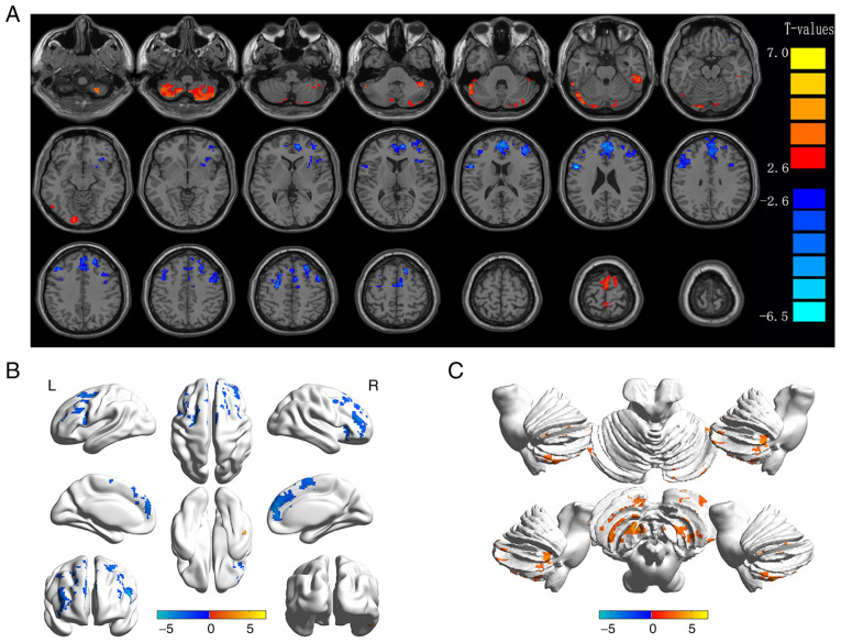
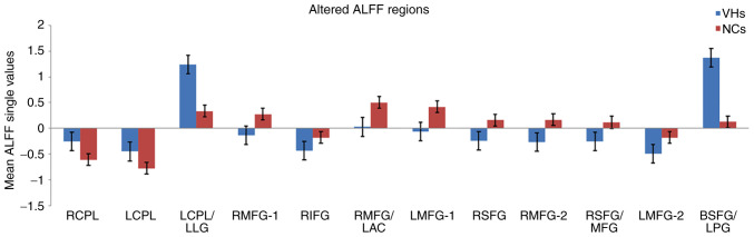
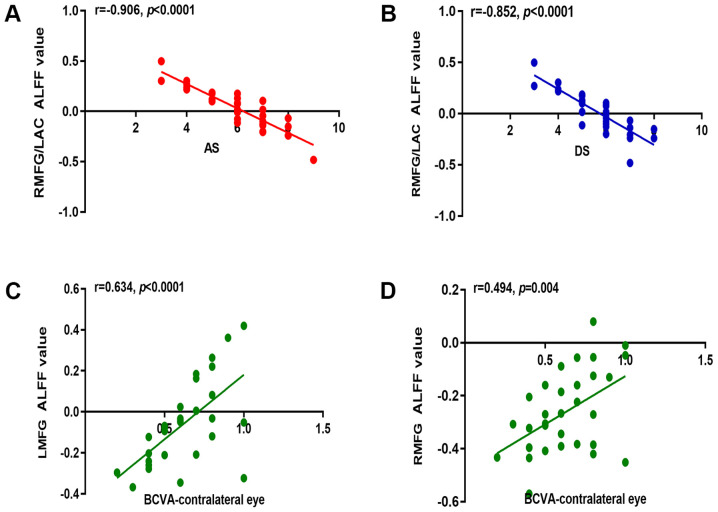
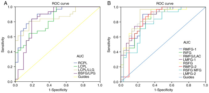
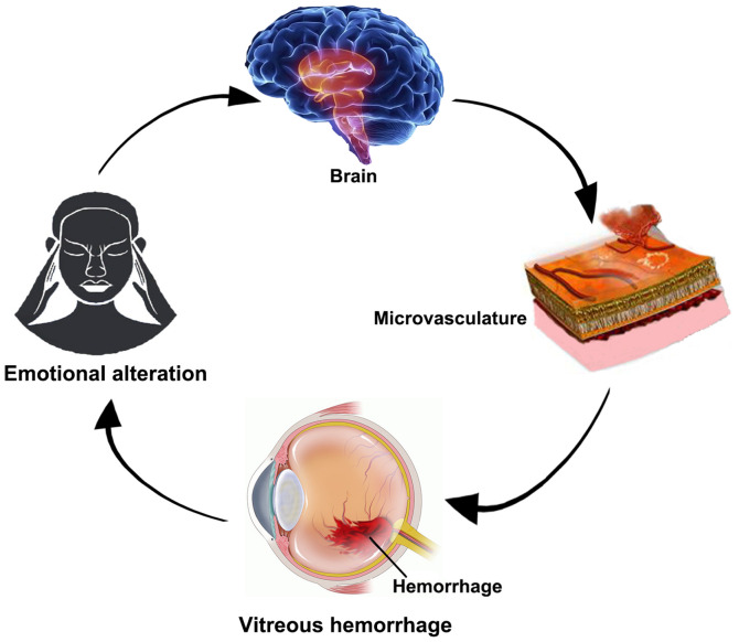
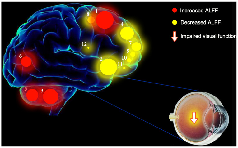
The aim of the present study was to assess the local character of spontaneous brain activity in type‑2 diabetic patients with vitreous hemorrhage (VH) and its relationship with clinical features via the amplitude of low‑frequency fluctuations (ALFF) method. A total of 31 subjects (15 females and 16 males) with type‑2 diabetic VH and 31 normal controls (NCs) with similar characteristics (sex, age and educational level) were recruited in the present study. All subjects underwent resting‑state functional magnetic resonance imaging scans. The local character of spontaneous brain activity was assessed using the ALFF method. The difference between the type‑2 diabetic patients with VH and NCs was determined using receiver operating characteristic curves. Pearson's correlation analysis was applied to evaluate the relationship between the mean ALFF values of specific brain areas and related clinical manifestations in type‑2 diabetic patients with VH. The ALFF values of type‑2 diabetic patients with VH were significantly increased in the right and left cerebellum posterior lobes, left cerebellum posterior lobe/left lingual gyrus and bilateral superior frontal gyrus/left postcentral gyrus, compared with those obtained for NCs (P<0.05). By contrast, these values were significantly decreased in the left and right middle frontal gyri, right medial frontal gyrus/left anterior cingulate, right inferior frontal gyrus, right superior frontal gyrus, right middle frontal gyrus, right superior frontal gyrus/middle frontal gyrus and left middle frontal gyrus of the former group compared with the NCs (P<0.05). Nevertheless, there was no significant association between the mean ALFF values and clinical characteristics in different brain areas. Unusual spontaneous activity occurred in multiple brain areas, which may suggest the neuropathological mechanisms of visual impairment in type‑2 diabetic patients with VH.

Keywords: vitreous hemorrhage; diabetes mellitus; amplitude of low-frequency; spontaneous brain activity.


Figures

Figure 1.
Figure 1.
Example of vitreous hemorrhage caused by PDR and RVO observed using a FC and FFA. PDR, proliferative diabetic retinopathy; RVO, retinal vein occlusion; FC, fundus camera; FFA, fluorescence fundus angiography.
Figure 2.
Figure 2.
Spontaneous brain activity in diabetic patients with VH vs. NCs. (A) Different ALFF regions between the VH and NC groups. (B) Differences of brain activity in the cerebrum. (C) Differences of brain activity in the cerebellum. Compared with HC, red represents the brain areas with increased ALFF, and blue represents the brain areas with decreased ALFF in patients with VH. P<0.01 for multiple comparisons using Gaussian Random Field theory (AlphaSim corrected; cluster >40 voxels; P<0.01). ALFF, amplitude of low-frequency fluctuation; VH, vitreous hemorrhage; NCs, normal controls; R, right; L, left.
Figure 3.
Figure 3.
Mean ALFF values between the VH and NC groups in the different regions of the brain. ALFF, amplitude of low-frequency fluctuation; VH, vitreous hemorrhage; NC, normal control; RCPL, right cerebellum posterior lobe; LCPL, left cerebellum posterior lobe; LLG, left lingual gyrus; RMFG, right middle frontal gyrus; RIFG, right inferior frontal gyrus; LAC, left anterior cingulate; LMFG, left middle frontal gyrus; RSFG, right superior frontal gyrus; MFG, middle frontal gyrus; BSFG/LPG, bilateral superior frontal gyrus/left postcentral gyrus.
Figure 4.
Figure 4.
Correlations between the mean ALFF value and behavioral performances in the different areas of the brain. (A) AS in the VH group displayed a negative correlation with the mean ALFF value of the RMFG/LAC (r=−0.906; P<0.0001). (B) DS in the VH group displayed a negative correlation with the mean ALFF value of the RMFG/LAC (r=−0.852; P<0.0001). In the VH group, BCVA of the contralateral eye displayed a positive correlation with the mean ALFF value of (C) the LMFG (r=0.634; P<0.0001) and (D) the RMFG (r=0.494; P=0.004). r, Pearson's correlation coefficient; ALFF, amplitude of low-frequency fluctuation; AS, anxiety score; RMFG, right middle frontal gyrus; LAC, left anterior cingulate; DS, depression score; BCVA, best-corrected visual acuity; LMFG, left middle frontal gyrus.
Figure 5.
Figure 5.
ROC curve analysis of the mean ALFF values of the altered brain regions in VH. (A) The areas under the ROC curve were: RCPL 0.893, (P<0.001; 95% CI, 0.814–0.972), LCPL 0.823 (P<0.001; 95% CI, 0.723–0.923), LCPL/LLG 0.856 (P<0.001; 95% CI, 0.761–0.951) and BSFG/LPG 0.839 (P<0.001; 95% CI, 0.732–0.945). (B) The areas under the ROC curve were: RMFG-1 0.847 (P<0.001; 95% CI, 0.748–0.946), RIFG 0.837 (P<0.001; 95% CI, 0.739–0.934), RMFG/LAC 0.890 (P<0.001; 95% CI, 0.807–0.972), LMFG-1 0.868 (P<0.001; 95% CI, 0.782–0.954), RSFG 0.864 (P<0.001; 95% CI, 0.773–0.954), RMFG-2 0.832 (P<0.001; 95% CI, 0.724–0.941), RSFG/MFG 0.781 (P<0.001; 95% CI, 0.666–0.897) and LMFG-2 0.850 (P<0.001; 95% CI, 0.754–0.947). ROC, receiver operating characteristic; ALFF, amplitude of low-frequency fluctuation; RCPL, right cerebellum posterior lobe; CI, confidence interval; LCPL, left cerebellum posterior lobe; LLG, left lingual gyrus; BSFG/LPG, bilateral superior frontal gyrus/left postcentral gyrus; RMFG, right middle frontal gyrus; RIFG, right inferior frontal gyrus; LAC, left anterior cingulate; LMFG, left middle frontal gyrus; RSFG, right superior frontal gyrus; MFG, middle frontal gyrus; AUC, area under the curve.
Figure 6.
Figure 6.
Relationship between magnetic resonance imaging and VH. Intracerebral hemorrhage may cause VH. Once VH occurs, it may affect the function of vision and lead to abnormal nerve activity in areas of the brain that are related to emotional processing. VH, vitreous hemorrhage.
Figure 7.
Figure 7.
Mean ALFF values of altered brain regions in the VH group. ALFF values of the following regions in diabetic patients with VH were increased to various extents compared with those obtained for NCs: (1) Cerebellum posterior lobe. L/lingual gyrus. L (BA 6; t=7.0388), (3) cerebellum posterior lobe. L (t=5.7220), (5) cerebellum posterior lobe. R (t=5.1342), and (6) cerebellum posterior lobe. L/lingual gyrus. L (BA 18; t=5.0742). By contrast, the ALFF values of the following regions were decreased: (2) Middle frontal gyrus. L (BA 10; t=−6.5002), (4) medial frontal gyrus. R (BA 9; t=−5.2619), (7) middle frontal gyrus. L (BA 10; t=−4.6431), (8) superior frontal gyrus. R (BA 6; t=−4.5000), (9) middle frontal gyrus. R (BA 10; t=−4.4753), (10) superior frontal gyrus. R /middle frontal gyrus (BA 10; t=−3.8568), (11) middle frontal gyrus. R (BA 10; t=−3.8469) and (12) inferior frontal gyrus. R (BA 45; t=−3.5367). The sizes of the spots denote the degree of quantitative changes in the ALFF values between the VH and NC groups. ALFF, amplitude of low-frequency fluctuations; VH, vitreous hemorrhage; NCs, normal controls; R, right; L, left; BA, Brodmann's area.

References

    1. Shaw JE, Sicree RA, Zimmet PZ. Global estimates of the prevalence of diabetes for 2010 and 2030. Diabetes Res Clin Pract. 2010;87:4–14. doi: 10.1016/j.diabres.2009.10.007. - DOI - PubMed
    1. Ong SR, Crowston JG, Loprinzi PD, Ramulu PY. Physical activity, visual impairment, and eye disease. Eye (Lond) 2018;32:1296–1303. doi: 10.1038/s41433-018-0081-8. - DOI - PMC - PubMed
    1. Spraul CW, Grossniklaus HE. Vitreous hemorrhage. Surv Ophthalmol. 1997;42:3–39. doi: 10.1016/S0039-6257(97)84041-6. - DOI - PubMed
    1. Yoonessi R, Hussain A, Jang TB. Bedside ocular ultrasound for the detection of retinal detachment in the emergency department. Acad Emerg Med. 2010;17:913–917. doi: 10.1111/j.1553-2712.2010.00809.x. - DOI - PubMed
    1. Cuevas P, Outeirino LA, Azanza C, Angulo J, Gimenez- Gallego G. Dramatic resolution of vitreous hemorrhage after an intravitreal injection of dobesilate. Mil Med Res. 2015;2:23. - PMC - PubMed