Observational Study
. 2020 Sep;44(3):838-848.
doi: 10.3892/or.2020.7669. Epub 2020 Jul 3.

Connective tissue growth factor produced by cancer‑associated fibroblasts correlates with poor prognosis in epithelioid malignant pleural mesothelioma

Affiliations
Observational Study

Connective tissue growth factor produced by cancer‑associated fibroblasts correlates with poor prognosis in epithelioid malignant pleural mesothelioma

Yuuki Ohara et al. Oncol Rep. 2020 Sep.

Abstract

Malignant mesothelioma is an aggressive neoplasm for which effective treatments are lacking. We often encounter mesothelioma cases with a profound desmoplastic reaction, suggesting the involvement of cancer‑associated fibroblasts (CAFs) in mesothelioma progression. While the roles of CAFs have been extensively studied in other tumors and have led to the view that the cancer stroma contains heterogeneous populations of CAFs, their roles in mesothelioma remain unknown. We previously showed that connective tissue growth factor (CTGF), a secreted protein, is produced by both mesothelioma cells and fibroblasts and promotes the invasion of mesothelioma cells in vitro. In this study, we examined the clinical relevance of CAFs in mesothelioma. Using surgical specimens of epithelioid malignant pleural mesothelioma, we evaluated the clinicopathological significance of the expression of α‑smooth muscle actin (αSMA), the most widely used marker of CAFs, the expression of CTGF, and the extent of fibrosis by immunohistochemistry and Elastica‑Masson staining. We also analyzed the expression of mesenchymal stromal cell‑ and fibroblast‑expressing Linx paralogue (Meflin; ISLR), a recently reported CAF marker that labels cancer‑restraining CAFs and differ from αSMA‑positive CAFs, by in situ hybridization. The extent of fibrosis and CTGF expression in mesothelioma cells did not correlate with patient prognosis. However, the expression of αSMA and CTGF, but not Meflin, in CAFs correlated with poor prognosis. The data suggest that CTGF+ CAFs are involved in mesothelioma progression and represent a potential molecular target for mesothelioma therapy.

Keywords: connective tissue growth factor; CTGF; cancer-associated fibroblasts; tumor microenvironment; malignant mesothelioma; mesenchymal stromal cell‑ and fibroblast‑expressing of a Linx paralogue; Meflin; ISLR; molecular target therapy.


Figures

Figure 1.
Figure 1.
Fibrotic and αSMA area indices in mesothelioma. (A) Calculation of the fibrotic area index using ImageJ. The image was obtained from mesothelioma in pleural fibrosis. (B) Analysis of Elastica-Masson staining. Fibrotic area indices for all cases are plotted as a histogram. (C) Calculation of the αSMA area index using ImageJ. DAB solution was used to stain αSMA, and HistoGreen was used to stain AE1/AE3. Mesothelioma cells were positive for AE1/AE3. The image was obtained from mesothelioma in pleural fibrosis. (D) Analysis of αSMA staining. The αSMA area indices for all cases are plotted as a histogram. (E) Fibrotic area index and patient prognosis based on Kaplan-Meier survival curves. There were no significant differences in prognosis based on the fibrotic area index (low, <40%; high, ≥40%). (F) αSMA expression and patient prognosis based on Kaplan-Meier survival curves. There was a significant difference (P=0.0262) in prognosis based on the αSMA area index (low, <20%; high, ≥20%). αSMA, α-smooth muscle actin; DAB, 3,3′-diaminobenzidine.
Figure 2.
Figure 2.
Immunohistochemical staining of CTGF. Both mesothelioma cells and CAFs were stained for CTGF. (A and B) Weak staining; score=1. The arrows in B indicate CAFs. (C and D) Moderate staining; score=2. (E and F) Strong staining; score=3. The arrows in E indicate mesothelioma cells. The arrows in F indicate CAFs. All images are shown at the same magnification. CAFs, cancer-associated fibroblasts; CTGF, connective tissue growth factor.
Figure 3.
Figure 3.
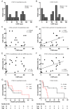
CTGF expression in CAFs correlates with mesothelioma patient prognosis. (A and B) Analysis of immunohistochemical staining of CTGF. The H-score of CTGF (CTGF score) for all cases is plotted as a histogram. (C) CTGF expression in mesothelioma cells and the Ki-67 index, indicating no significant differences. (D) CTGF expression in CAFs and the Ki-67 index, indicating a positive correlation (r=0.533, P=0.0107; Spearman's correlation test). (E) CTGF expression in mesothelioma cells and the αSMA area index, indicating no significant differences. (F) CTGF expression in CAFs and the αSMA area index, indicating a positive correlation (r=0.502, P=0.0172; Spearman's correlation test). (G) CTGF expression in mesothelioma cells and patient prognosis based on Kaplan-Meier survival curves. CTGF scores were modified by the AE1/AE3 area index values. There were no significant differences in prognosis based on modified CTGF scores (low, <10; high, ≥10). (H) CTGF expression in CAFs and patient prognosis based on Kaplan-Meier survival curves. CTGF scores were modified by the αSMA area index values. There was a significant difference (P=0.0186) in prognosis based on modified CTGF score (low, <30; high, ≥30). αSMA, α-smooth muscle actin; CAFs, cancer-associated fibroblasts; CTGF, connective tissue growth factor.
Figure 4.
Figure 4.
Meflin expression in mesothelioma. (A) RNA ISH of Meflin. DAB solution was used to stain αSMA, and HistoGreen was used to stain AE1/AE3. Mesothelioma cells were positive for AE1/AE3. CAFs and vessels were positive for αSMA. More Meflin-positive (Meflin+) CAFs were observed in the αSMA-negative area (top), which is the invasive front of mesothelioma. Less Meflin+ CAFs were observed in the αSMA-high area (bottom), which is the proximal side of mesothelioma. The merged images of αSMA + AE1/AE3 and Meflin were obtained using ImageJ software. Meflin-positive area is indicated by red color in the merged images. All images are shown at the same magnification. (B) Analysis of RNA ISH data for Meflin. The proportions of the Meflin+ CAFs for all cases are plotted as a histogram. (C) Meflin expression in CAFs and patient prognosis based on Kaplan-Meier survival curves. There was no significant difference in prognosis based on the proportions of the Meflin+ CAFs (low, <10%; high, ≥10%). Gehan-Breslow-Wilcoxon test was used for analysis. αSMA, α-smooth muscle actin; CAFs, cancer-associated fibroblasts; H&E, hematoxylin and eosin; ISH, in situ hybridization; Meflin, mesenchymal stromal cell- and fibroblast-expressing Linx paralogue.
Figure 5.
Figure 5.
Fibroblasts in mesothelial carcinogenesis. Previous studies suggest the existence of three phenotypes of fibroblasts: Quiescent, wound healing-associated, and mesothelioma cell-educated. Asbestos fibers, which contain iron as a component, can directly induce reactive oxygen species (ROS) generation via catalysis of the Fenton reaction by iron on the surface. Macrophages phagocytose asbestos fibers and form granulomas. These macrophages can also produce ROS. ROS can induce quiescent fibroblasts to differentiate into myofibroblasts. These αSMA+ fibroblasts can contribute to carcinogenesis by secreting CTGF and cytokines. Fibroblasts educated by mesothelioma cells express CTGF and have protumorigenic roles. Meflin+ fibroblasts may have antitumorigenic roles. αSMA, α-smooth muscle actin; CTGF, connective tissue growth factor; Meflin, mesenchymal stromal cell- and fibroblast-expressing Linx paralogue.

References

    1. International Agency for Research on Cancer, corp-author. A Review of Human Carcinogens; Part C: Arsenic. Metals; Fibres, and Dusts, Lyon, France: 2012. WHO. Asbestos (chrysotile, amosite, crocidolite, tremolite, actinolite, and anthophyllite). IARC Monographs on the Evaluation of Carcinogenic Risks to Humans Volume 100C; pp. 219–309.
    1. Pavlisko EN, Sporn TA. Pathology of asbestos-associated diseases. 3rd. Springer; Berlin: 2014. Mesothelioma; pp. 81–140. - DOI
    1. Rusch VW, Giroux D, Kennedy C, Ruffini E, Cangir AK, Rice D, Pass H, Asamura H, Waller D, Edwards J, et al. Initial analysis of the international association for the study of lung cancer mesothelioma database. J Thorac Oncol. 2012;7:1631–1639. doi: 10.1097/JTO.0b013e31826915f1. - DOI - PubMed
    1. Donaldson K, Murphy FA, Duffin R, Poland CA. Asbestos, carbon nanotubes and the pleural mesothelium: A review of the hypothesis regarding the role of long fibre retention in the parietal pleura, inflammation and mesothelioma. Part Fibre Toxicol. 2010;7:5. doi: 10.1186/1743-8977-7-5. - DOI - PMC - PubMed
    1. Nagai H, Toyokuni S. Biopersistent fiber-induced inflammation and carcinogenesis: Lessons learned from asbestos toward safety of fibrous nanomaterials. Arch Biochem Biophys. 2010;502:1–7. doi: 10.1016/j.abb.2010.06.015. - DOI - PubMed

Publication types

MeSH terms