. 2020 Nov;30(6):1087-1101.
doi: 10.1111/bpa.12888. Epub 2020 Aug 14.

Loss of capillary pericytes and the blood-brain barrier in white matter in poststroke and vascular dementias and Alzheimer's disease

Affiliations

Loss of capillary pericytes and the blood-brain barrier in white matter in poststroke and vascular dementias and Alzheimer's disease

Ren Ding et al. Brain Pathol. 2020 Nov.

Abstract

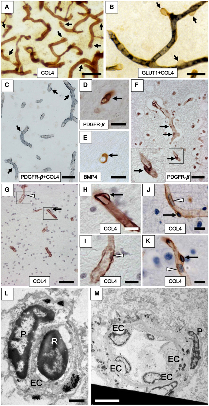
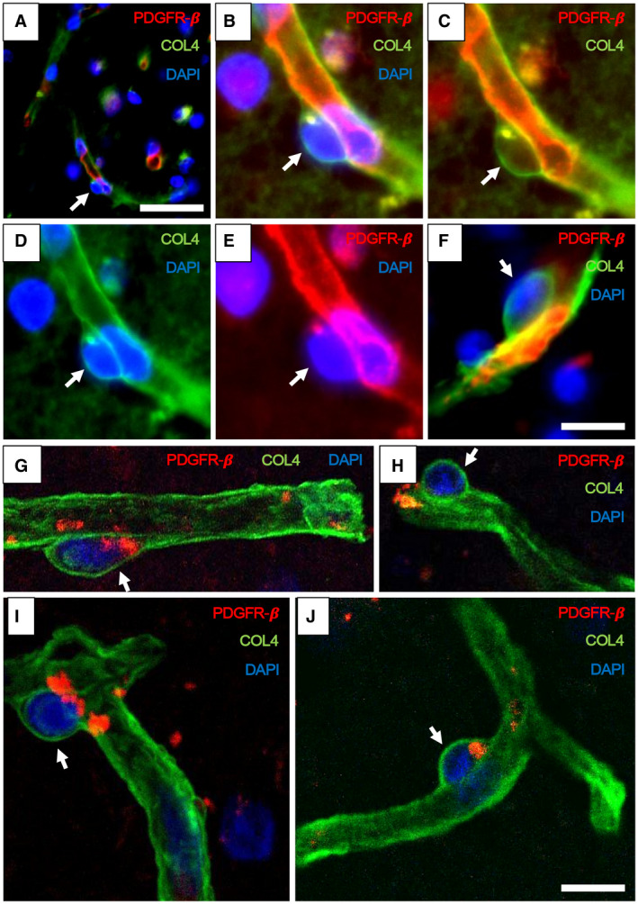
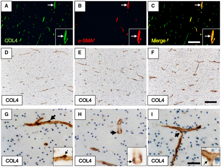
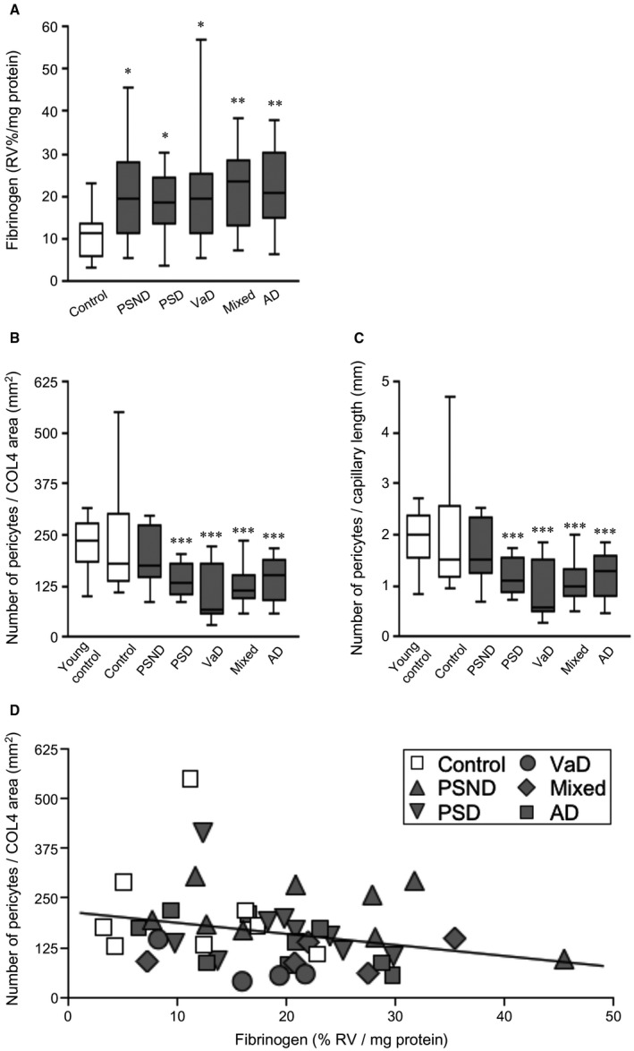
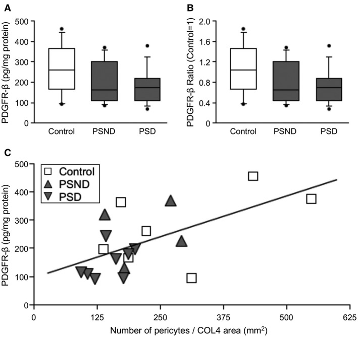
White matter (WM) disease is associated with disruption of the gliovascular unit, which involves breach of the blood-brain barrier (BBB). We quantified pericytes as components of the gliovascular unit and assessed their status in vascular and other common dementias. Immunohistochemical and immunofluorescent methods were developed to assess the distribution and quantification of pericytes connected to the frontal lobe WM capillaries. Pericytes with a nucleus were identified by collagen 4 (COL4) and platelet-derived growth factor receptor-β (PDGFR-β) antibodies with further verification using PDGFR-β-specific ELISA. We evaluated a total of 124 post-mortem brains from subjects with post-stroke dementia (PSD), vascular dementia (VaD), Alzheimer's disease (AD), AD-VaD (Mixed) and post-stroke non-demented (PSND) stroke survivors as well as normal aging controls. COL4 and PDGFR-β reactive pericytes adopted the characteristic "crescent" or nodule-like shapes around capillary walls. We estimated densities of pericyte somata to be 225 ±38 and 200 ±13 (SEM) per COL4 mm2 area or 2.0 ± 0.1 and 1.7 ± 0.1 per mm capillary length in young and older aging controls. Remarkably, WM pericytes were reduced by ~35%-45% in the frontal lobe of PSD, VaD, Mixed and AD subjects compared to PSND and controls subjects (P < 0.001). We also found pericyte numbers were correlated with PDGFR-β reactivity in the WM. Our results first demonstrate a reliable method to quantify COL4-positive pericytes and then, indicate that deep WM pericytes are decreased across different dementias including PSD, VaD, Mixed and AD. Our findings suggest that downregulation of pericytes is associated with the disruption of the BBB in the deep WM in several aging-related dementias.

Keywords: cerebral capillary; collagen type IV; dementia; pericytes; platelet-derived growth factor receptor; vascular dementia; white matter.


Conflict of interest statement

The authors have no disclosures or conflicts of interest in relation to this manuscript.

Figures

Figure 1
Figure 1
Cerebral capillaries with pericytes in the frontal cortex and WM of aging subjects. A–F. Pericytes evident as “bumps” or “crescent” shaped cells in the capillary network immunostained with antibodies to COL4 (A, G, H, I, J and K), GLUT1 (blue‐black) and COL4 (brown) (B), PDGFR‐β (brown) and COL4 (blue/gray) (C), PDGFR‐β (D, F) and BMP4 (E). Arrows show location of the pericyte cell bodies along the capillaries. G–K. Pericyte soma (black arrows in G, H, J and K) and endothelial cell (white arrowhead in G and I) and other types of cells including erythrocytes observed within the lumen (white arrowhead in J and K) in the frontal WM capillaries of the Baboon (GI) and human (J and K). L,M. Transmission electron microscope images show pericyte cell body (P) on a WM capillary (L) and precapillary/small arteriole (M) in cross‐section from 65‐year‐old male subject. The marked differential localization between the pericyte and endothelial cell (EC) and the lumen with an erythrocyte (R for red blood cell) can be noted. L–M also assure assessment of non‐capillary pericytes or EC or smooth muscle cell nuclei were not included in the pericyte counts. Images in A and B were from 20 µm thick sections, whereas those from C to I were from 10 µm sections. Length density of frontal cortical capillaries (A) was greater than WM (B). Magnification bars: A, G = 50 µm; C, F = 30 µm; B, D, E = 20 µm; H, I, J, K = 10 µm; L = 1 µm; M = 5 µm.
Figure 2
Figure 2
Pericyte soma on WM capillaries demonstrated by immunofluorescence. A–F. PDGFR‐β (red) and COL4 (green) immunofluorescence staining with nuclei (DAPI) (blue). A,B. Low‐ and high‐power images showing capillary segments (arrowheads) with overlapping PDGFR‐β (red), COL4 (green) and DAPI (blue). C. Same vessel segment as B with PDGFR‐β (red) and COL4 (green); D. COL4 (green) and DAPI (blue); E. PDGFR‐β (red) and DAPI (blue). F. Another capillary segment with PDGFR‐β (red), COL4 (green) and DAPI (blue) clearly showing pericyte cell body. G–J. Images taken by a confocal microscope showing pericyte cell bodies (arrows) with nuclear stain (DAPI). Capillaries and pericyte processes are revealed by COL4 and PDGFR‐β (red) immunoreactivities. Magnification bars: A = 50 µm, F, J = 10 µm.
Figure 3
Figure 3
Quantification methods used for determining density of pericytes associated with capillaries. A–C. Immunofluorescent staining of WM capillaries and arterioles. A. COL4 (green). B. α‐SMA (red). C. merged image of A and B. Quantification of pericytes was performed on COL4‐positive but α‐SMA‐negative capillary profiles. Each inset showing an arteriole double‐positive for COL4 and α‐SMA (white arrow) at higher magnification. D–I. Pericyte cell bodies revealed by COL4 immunostaining and hematoxylin nuclear counterstain as crescent shapes or as bumps on capillaries of the WM. Pericytes cell bodies are evident in longitudinal (G) and transverse profiles (H) as well as at intersections (I) of capillaries. G–I. Each inset showing a pericyte (arrow) at higher power. Magnification bars: C, F = 200 µm; I = 50 µm.
Figure 4
Figure 4
Quantification of WM pericytes in different dementias compared to normal aging controls. A. Fibrinogen reactivity in frontal WM in control, PSND and different dementia groups. Fibrinogen reactivity measured by ELISA in WM extracts was increased in PSND and all the dementia groups compared to controls (*P < 0.05; **P < 0.01). B,C. Box plots show pericyte numbers per COL4 area (mm2) (B) and per unit (mm) capillary length (C). Pericyte numbers were decreased in PSD compared to PSND subjects (***P < 0.001). They were also decreased in all the dementia types compared to controls and young controls. (***P < 0.001, one‐way ANOVA, n = 8–12 cases). Data generated from counts of pericytes per case comprising 110 to 385 cells per each group derived from 82 to 382 separate images. D. Correlation of fibrinogen reactivity (% RV, relative values) with pericyte number per COL4 area (mm2) (Pearson’s r = −0.3, P = 0.057). Demographic details of the subjects are given in Table 1.
Figure 5
Figure 5
Quantification of PDGFR‐β reactivity by ELISA in the frontal WM. A,B. Box plots show relative PDGFR‐β reactivity in extracts of the WM from PSD subjects compared to PSND and normal controls. A. Plots show PDGFR‐β value (pg/mg protein). B. Plots represent PDGFR‐β ratio standardised to control (control = 1). C. Correlation of PDGFR‐β reactivity by ELISA and pericyte number per COL4 area in frontal WM. PDGFR‐β detected by ELISA was correlated with pericyte number per COL4 area (Pearson’s r = 0.6, P = 0.007).
Figure 6
Figure 6
Integrity of the BBB and pericyte numbers in the frontal WM in a nonhuman primate model of cerebral hypoperfusion. A. Box plots showing mean vascular density (% COL4 stained area) in the frontal WM of adult baboons subjected to 3VO. Each time point represents mean of n = 4–8 animals and the results were computed from both hemispheres. There were no differences between the right and left sides. Microvascular density was not changed after 3VO compared to Sham operated animals (P > 0.05). B. Quantification of pericytes density and fibrinogen reactivity in the frontal WM of adult baboons subjected to 3VO. There was a high variation in fibrinogen immunoreactivity over survival time (P < 0.01). Fibrinogen immunoreactivity was the highest at 14 days after 3VO compared to Sham, 1 and 28 days after 3VO groups (*P < 0.05), concomitant with relatively lower pericyte density at 14 days.

References

    1. Akinyemi RO, Allan LM, Oakley A, Kalaria RN (2017) Hippocampal neurodegenerative pathology in post‐stroke dementia compared to other dementias and aging controls. Front Neurosci 11:717. - PMC - PubMed
    1. Allan LM, Rowan EN, Firbank MJ, Thomas AJ, Parry SW, Polvikoski TM et al (2011) Long term incidence of dementia, predictors of mortality and pathological diagnosis in older stroke survivors. Brain 134(Pt 12):3716–3727. - PMC - PubMed
    1. Andrae J, Gallini R, Betsholtz C (2008) Role of platelet‐derived growth factors in physiology and medicine. Genes Dev 22:1276–1312. - PMC - PubMed
    1. Armulik A, Genove G, Betsholtz C (2011) Pericytes: developmental, physiological, and pathological perspectives, problems, and promises. Dev Cell 21:193–215. - PubMed
    1. Baradaran H, Mtui EE, Richardson JE, Delgado D, Dunning A, Marshall RS et al (2016) White matter diffusion abnormalities in carotid artery disease: a systematic review and meta‐analysis. J Neuroimaging 26:481–488. - PubMed

Publication types

MeSH terms

Substances