

Profiling neovascular age-related macular degeneration choroidal neovascularization lesion response to anti-vascular endothelial growth factor therapy using SSOCTA
- PMID: 32706171
- PMCID: PMC7984400
- DOI: 10.1111/aos.14554
Profiling neovascular age-related macular degeneration choroidal neovascularization lesion response to anti-vascular endothelial growth factor therapy using SSOCTA
Abstract
Purpose: To identify the changes in distinct vascular parameters of choroidal neovascularization (CNV) in eyes with treatment-naïve neovascular age-related macular degeneration (nAMD) during the primary response to anti-VEGF therapy using aflibercept.
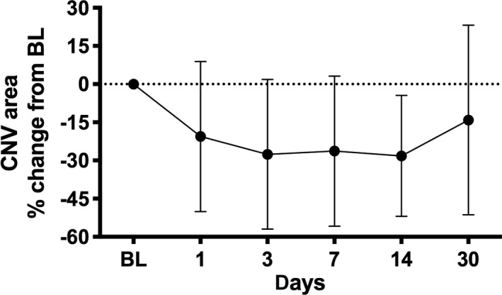
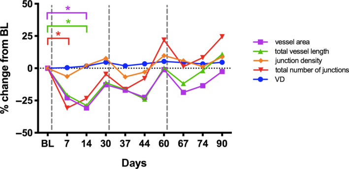
Methods: Patients were prospectively followed during the first 3 months according to a standardized protocol with mandatory visits at days 7 and 14 after each anti-VEGF treatment up to day 90. Fourteen eyes were seen in addition at days 1 and 3 post-initial injection. Aflibercept was administered at baseline (BL), day 30 and 60. 6 × 6mm SSOCTA (PlexElite, Zeiss) images were acquired. Using the semi-automated AngioTool, CNV area, vessel area, vessel density (VD), the number of junctions, junctions density, total vessel length, average vessel length, total number of endpoints and lacunarity were assessed.
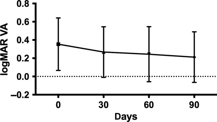
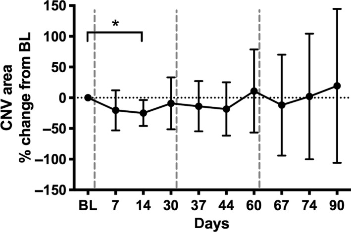
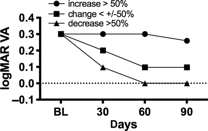
Results: Thirty-two consecutive patients presenting with treatment-naïve, SSOCTA-positive CNV lesions were included. Close follow-up showed a characteristic neovascular response curve with a dynamic decrease in lesion size within days and a reactive increase following 2 weeks after initial treatment. An undulating pattern was seen for all neovascular parameters except for vascular density, with variable statistical significance. Due to a flattening of the therapeutic response as early as after the second treatment, CNV lesion size and most of the related parameters had an increase in activity above baseline values at the end of the loading phase. Lesion size was the leading feature of reactivation by a mean increase of 19.3% after three monthly aflibercept injections. Subgroup analysis based on lesion size revealed a significant correlation between best-corrected visual acuity and quantitative change in lesion size over time, but not baseline size.
Conclusions: Using SSOCTA, a morphologic neovascular response pattern can be identified in anti-VEGF treatment of CNV. A synchronized early decrease and consecutive reactivation in a large spectrum of neovascular biomarkers including size and internal structure are visualized in a qualitative and quantitative manner. SSOCTA analyses allow new insights in CNV morphology changes and therapeutic response.
Keywords: SSOCTA; biomarker; choroidal neovascularization; loading phase; neovascular age-related macular degeneration.
© 2020 The Authors. Acta Ophthalmologica published by John Wiley & Sons Ltd on behalf of Acta Ophthalmologica Scandinavica Foundation.
Figures
References
-
- Babiuch AS, Uchida A, Figueiredo N et al. (2019): Impact of optical coherence tomography angiography review strategy on detection of choroidal neovascularization. Retina 40: 672–678. - PubMed
-
- Bogunovic H, Waldstein SM, Schlegl T et al. (2017): Prediction of anti‐VEGF treatment requirements in neovascular AMD using a machine learning approach. Invest Ophthalmol Vis Sci 58: 3240–3248. - PubMed
-
- Brown DM, Kaiser PK, Michels M et al. (2006): Ranibizumab versus verteporfin for neovascular age‐related macular degeneration. N Engl J Med 355: 1432–1444. - PubMed
-
- de Carlo TE, Bonini Filho MA, Chin AT et al. (2015): Spectral‐domain optical coherence tomography angiography of choroidal neovascularization. Ophthalmology 122: 1228–1238. - PubMed
-
- Carnevali A, Sacconi R, Querques L et al. (2018): Natural history of treatment‐naive quiescent choroidal neovascularization in age‐related macular degeneration using OCT angiography. Ophthalmol Retina 2: 922–930. - PubMed
MeSH terms
Substances
LinkOut - more resources
Full Text Sources
