Practice Guideline

Diagnosis of Hypersensitivity Pneumonitis in Adults. An Official ATS/JRS/ALAT Clinical Practice Guideline

Ganesh Raghu et al. Am J Respir Crit Care Med. .

Erratum in

Abstract

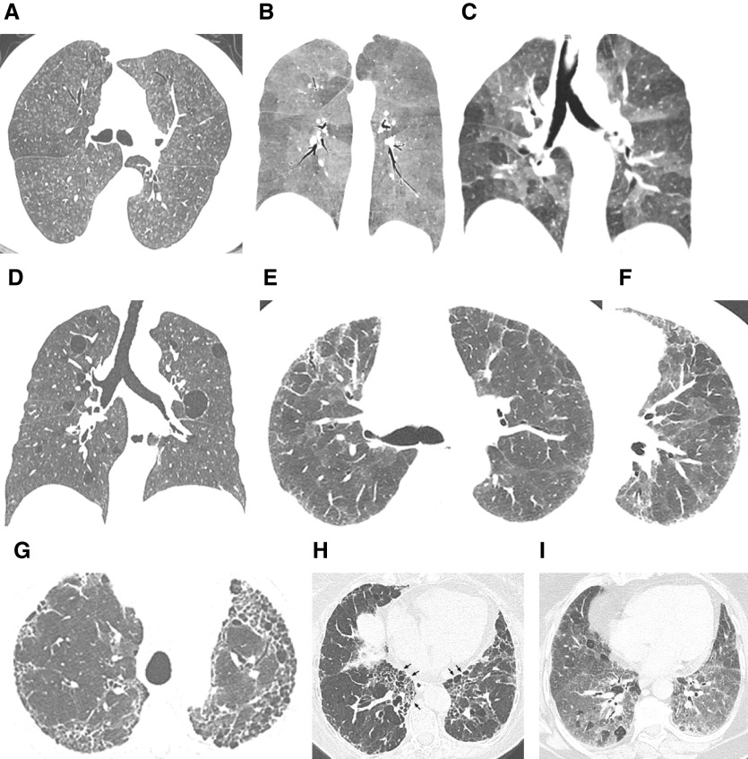
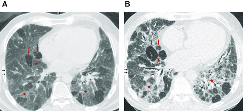
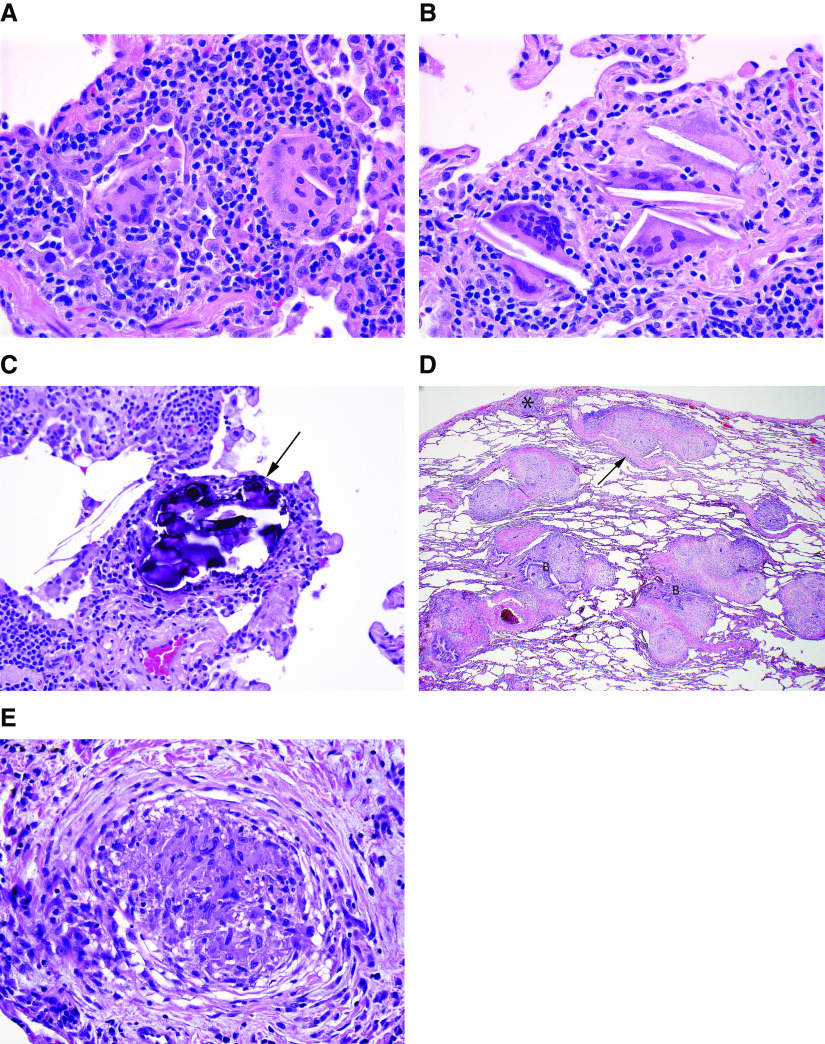
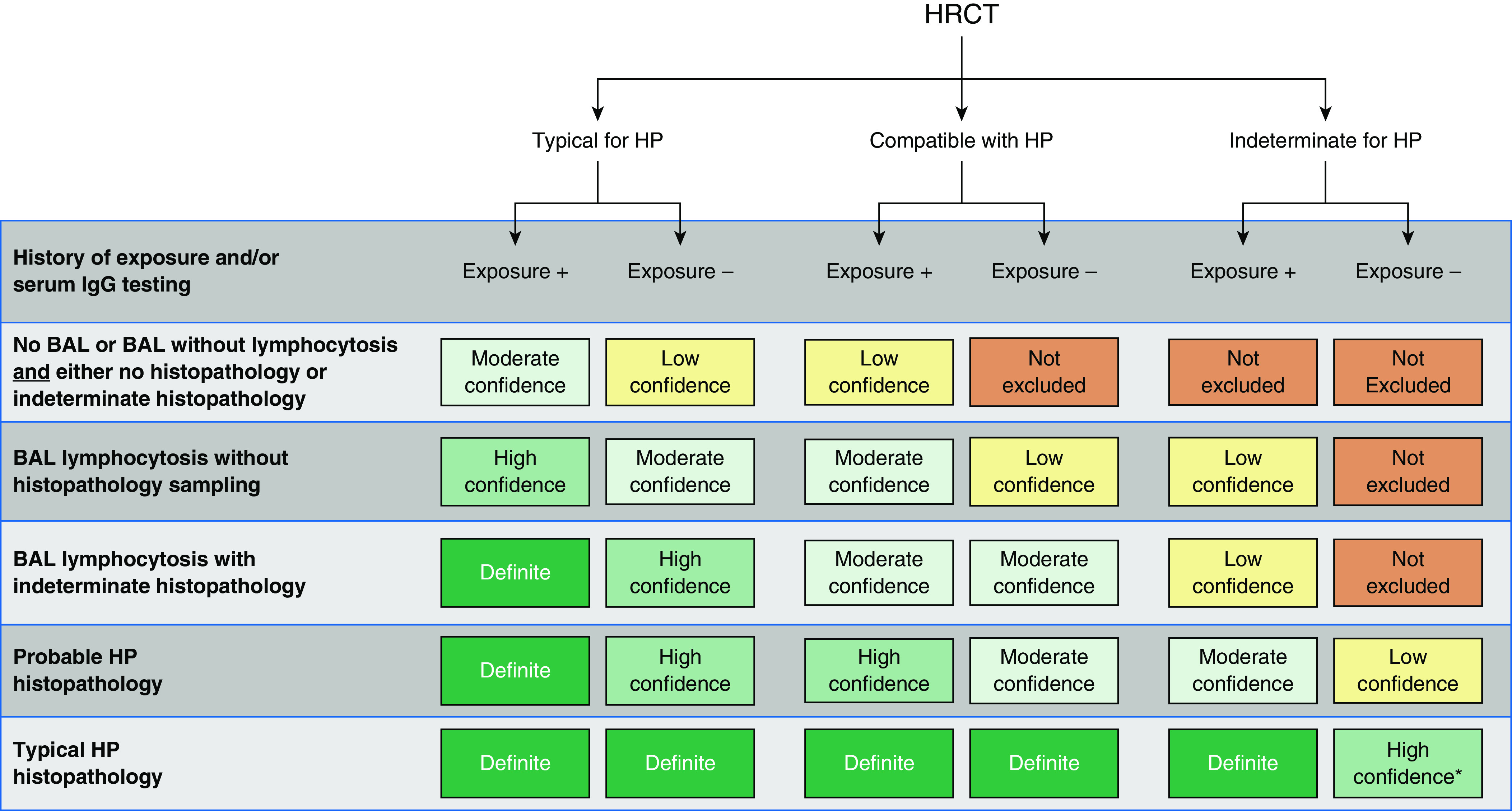
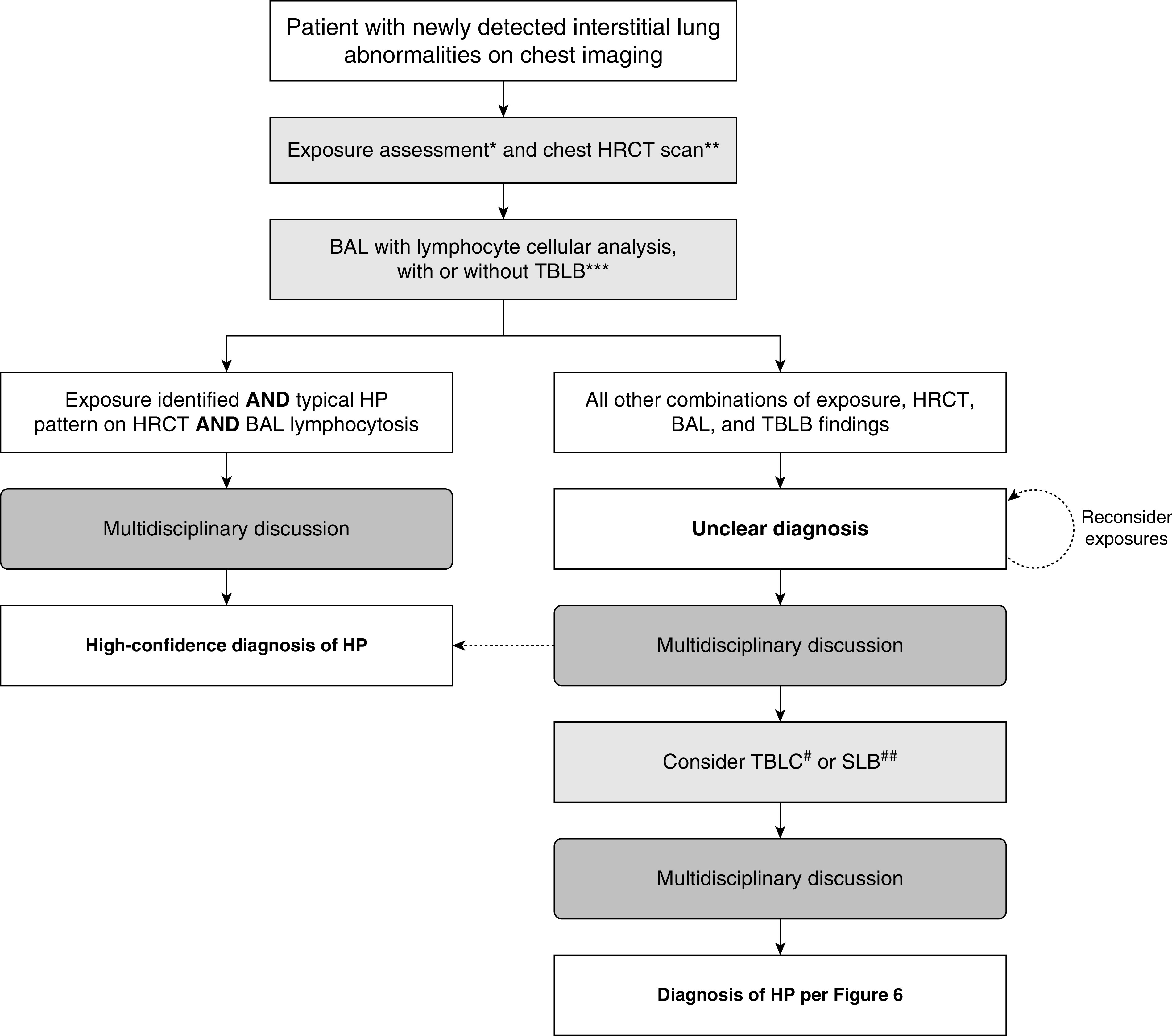
Background: This guideline addresses the diagnosis of hypersensitivity pneumonitis (HP). It represents a collaborative effort among the American Thoracic Society, Japanese Respiratory Society, and Asociación Latinoamericana del Tórax.Methods: Systematic reviews were performed for six questions. The evidence was discussed, and then recommendations were formulated by a multidisciplinary committee of experts in the field of interstitial lung disease and HP using the GRADE (Grading of Recommendations, Assessment, Development, and Evaluation) approach.Results: The guideline committee defined HP, and clinical, radiographic, and pathological features were described. HP was classified into nonfibrotic and fibrotic phenotypes. There was limited evidence that was directly applicable to all questions. The need for a thorough history and a validated questionnaire to identify potential exposures was agreed on. Serum IgG testing against potential antigens associated with HP was suggested to identify potential exposures. For patients with nonfibrotic HP, a recommendation was made in favor of obtaining bronchoalveolar lavage (BAL) fluid for lymphocyte cellular analysis, and suggestions for transbronchial lung biopsy and surgical lung biopsy were also made. For patients with fibrotic HP, suggestions were made in favor of obtaining BAL for lymphocyte cellular analysis, transbronchial lung cryobiopsy, and surgical lung biopsy. Diagnostic criteria were established, and a diagnostic algorithm was created by expert consensus. Knowledge gaps were identified as future research directions.Conclusions: The guideline committee developed a systematic approach to the diagnosis of HP. The approach should be reevaluated as new evidence accumulates.

Keywords: fibrotic hypersensitivity pneumonitis; hypersensitivity pneumonitis; interstitial lung disease; nonfibrotic hypersensitivity pneumonitis; pulmonary fibrosis.


Figures

Figure 1.
Figure 1.
“Typical hypersensitivity pneumonitis (HP)” and “compatible-with-HP” high-resolution computed tomography patterns. The nonfibrotic typical HP pattern is characterized by (A) centrilobular nodules, (B) mosaic attenuation on an inspiratory scan, and (C) air trapping on an expiratory scan. (D) The nonfibrotic compatible-with-HP pattern is exemplified by uniform and subtle ground-glass opacity and cysts. The fibrotic typical HP pattern consists of (E) coarse reticulation and minimal honeycombing in a random axial distribution with no zonal predominance in association with (F) small airway disease. The fibrotic compatible-with-HP pattern varies in the patterns and/or distribution of lung fibrosis (e.g., basal and subpleural predominance, [G] upper-lung-zone predominance, [H] central [or peribronchovascular] predominance [arrows], or [I] fibrotic ground-glass attenuation seen alone or in association with small airway disease). The fibrotic indeterminate-for-HP pattern includes the usual interstitial pneumonia pattern, nonspecific interstitial pneumonia pattern, organizing pneumonia–like pattern, or truly indeterminate findings.
Figure 2.
Figure 2.
Three-density pattern. High-resolution computed tomography (A) inspiratory and (B) expiratory images from a patient with hypersensitivity pneumonitis demonstrating the three different densities: high attenuation (ground-glass opacity) (red stars), lucent lung (regions of decreased attenuation and decreased vascular sections) (red arrows), and normal lung (black arrows), which are sharply demarcated from each other.
Figure 3.
Figure 3.
Surgical lung biopsy specimen from a patient with nonfibrotic hypersensitivity pneumonitis (HP). (A) Low-magnification photomicrograph showing preservation of lung architecture and a cellular chronic interstitial pneumonia that is accentuated around bronchioles (asterisks). Magnification, 20×. (B) Higher-magnification photomicrograph showing expansion of distal acinar and peribronchiolar interstitium by a cellular infiltrate of mononuclear inflammatory cells. Magnification, 88×. (C) Photomicrograph showing a cellular bronchiolitis in which the peribronchiolar interstitium is expanded by cellular infiltrate, predominantly comprising lymphocytes without lymphoid aggregates or follicles. Magnification, 108×. (D) Higher-magnification view of airway illustrated in C, demonstrating a poorly formed nonnecrotizing granuloma (arrow) characteristic of HP comprising loose clusters of epithelioid cells (macrophages). Magnification, 400×. (E) High-magnification photomicrograph illustrating another poorly formed nonnecrotizing granuloma (arrows) in the same biopsy specimen from a patient with nonfibrotic HP. Magnification, 264×. Hematoxylin and eosin staining was used.
Figure 4.
Figure 4.
(AC) Poorly formed granulomas characteristic of hypersensitivity pneumonitis (HP) contrasted with (D and E) well-formed granulomas more typical of sarcoidosis. (A) High-magnification photomicrograph illustrating isolated multinucleated giant cells in a surgical lung biopsy specimen from a patient with nonfibrotic HP. Magnification, 400×. (B) Another photomicrograph illustrating giant cells in a patient with HP. These giant cells are distinguished by cytoplasmic cholesterol-like clefts, a nonspecific but common finding. Magnification, 400×. (C) In this high-magnification photomicrograph of a surgical lung biopsy specimen, the giant cells are largely obscured by cytoplasmic Schaumann bodies (arrow), another nonspecific but characteristic feature of the granulomatous response in HP. Magnification, 400×. (D) Low-magnification photomicrograph of surgical lung biopsy specimen from a patient with sarcoidosis showing characteristic “lymphangitic” distribution, in which the granulomas are limited to the interstitium and involve visceral pleura (asterisk), interlobular septa (arrow), and bronchovascular bundles. Magnification, 20×. (E) High-magnification photomicrograph showing a well-formed nonnecrotizing granuloma in a surgical lung biopsy specimen from a patient with sarcoidosis. The well-circumscribed, tight cluster of epithelioid cells (macrophages) is affiliated with a characteristic pattern of circumferential lamellar fibrosis. Magnification, 400×. Hematoxylin and eosin staining was used. B = bronchovascular bundle.
Figure 5.
Figure 5.
Photomicrographs of surgical lung biopsy specimens from two different sites in a patient with fibrotic hypersensitivity pneumonitis. (A) Scanning magnification view showing multiple sections of a right-lower-lobe biopsy specimen. There is patchy fibrosis with architectural distortion, a combination of findings that resembles usual interstitial pneumonia. Magnification, 6×. (B) Low-magnification photomicrograph showing one of the sections illustrated in A, characterized by a pattern of patchy fibrosis with subpleural honeycomb change that resembles usual interstitial pneumonia. Magnification, 17×. (C) Higher-magnification view showing expansion of the peribronchiolar interstitium by a cellular infiltrate of mononuclear inflammatory cells (upper left) and isolated Schaumann bodies (arrows) at the edge of the biopsy specimen. Magnification, 46×. (D) High-magnification photomicrograph showing one of the isolated Schaumann bodies illustrated in C. Magnification, 400×. (E) Photomicrograph from another section illustrated in A showing an isolated Schaumann body (arrow) in the fibrotic peribronchiolar interstitium. Magnification, 63×. (F) Low-magnification photomicrograph of a right-middle-lobe biopsy specimen from the same patient showing features more closely resembling nonfibrotic hypersensitivity pneumonitis. There is a more cellular chronic interstitial pneumonia accentuated around bronchioles with scattered calcified Schaumann bodies (arrows) marking isolated multinucleated giant cells. Magnification, 43×. Hematoxylin and eosin staining was used. B = bronchiole.
Figure 6.
Figure 6.
Hypersensitivity pneumonitis diagnosis based on incorporation of imaging, exposure assessment, BAL lymphocytosis, and histopathological findings. All confidence levels are subject to multidisciplinary discussion. *Confidence may increase to “definite” if the pathologist’s conclusion persists after reevaluation in the context of additional clinical information or an expert second opinion on histopathology. HP = hypersensitivity pneumonitis; HRCT = high-resolution computed tomography.
Figure 7.
Figure 7.
Algorithm for the diagnostic evaluation of possible hypersensitivity pneumonitis (HP). Specific features are described for all steps of the algorithm in the corresponding sections of the manuscript. A provisional diagnosis may be adequate in patients for whom the differential diagnosis has been sufficiently narrowed such that further investigations are unlikely to alter management, when invasive testing has unacceptable risks, or when such tests are declined by the patient. *Exposure assessment includes a thorough clinical history and/or serum IgG testing against potential antigens associated with HP and/or, in centers with the appropriate expertise and experience, specific inhalational challenge testing as described in References , , , and . **High-resolution computed tomography should be performed using the technique described in Table 3 and then reviewed with a thoracic radiologist. ***Transbronchial lung biopsy is suggested for patients with potential nonfibrotic HP (see question 4, recommendation 1). #TBLC is suggested for patients with potential nonfibrotic HP, depending on local expertise (see question 5, recommendation 2). ##SLB is infrequently considered in patients with nonfibrotic HP. HRCT = high-resolution computed tomography; SLB = surgical lung biopsy; TBLB = transbronchial lung biopsy; TBLC = transbronchial lung cryobiopsy.

Comment in

References

    1. Lacasse Y, Selman M, Costabel U, Dalphin JC, Ando M, Morell F, et al. HP Study Group. Clinical diagnosis of hypersensitivity pneumonitis. Am J Respir Crit Care Med. 2003;168:952–958. - PubMed
    1. Elicker BM, Jones KD, Henry TS, Collard HR. Multidisciplinary approach to hypersensitivity pneumonitis. J Thorac Imaging. 2016;31:92–103. - PubMed
    1. Churg A, Muller NL, Flint J, Wright JL. Chronic hypersensitivity pneumonitis. Am J Surg Pathol. 2006;30:201–208. - PubMed
    1. Fink JN, Ortega HG, Reynolds HY, Cormier YF, Fan LL, Franks TJ, et al. Needs and opportunities for research in hypersensitivity pneumonitis. Am J Respir Crit Care Med. 2005;171:792–798. - PubMed
    1. Schuyler M, Cormier Y. The diagnosis of hypersensitivity pneumonitis. Chest. 1997;111:534–536. - PubMed

Publication types

MeSH terms

Substances