Case Reports
. 2020 Aug 18;324(7):663-673.
doi: 10.1001/jama.2020.13719.

Presence of Genetic Variants Among Young Men With Severe COVID-19

Affiliations
Case Reports

Presence of Genetic Variants Among Young Men With Severe COVID-19

Caspar I van der Made et al. JAMA. .

Abstract

Importance: Severe coronavirus disease 2019 (COVID-19) can occur in younger, predominantly male, patients without preexisting medical conditions. Some individuals may have primary immunodeficiencies that predispose to severe infections caused by severe acute respiratory syndrome coronavirus 2 (SARS-CoV-2).

Objective: To explore the presence of genetic variants associated with primary immunodeficiencies among young patients with COVID-19.

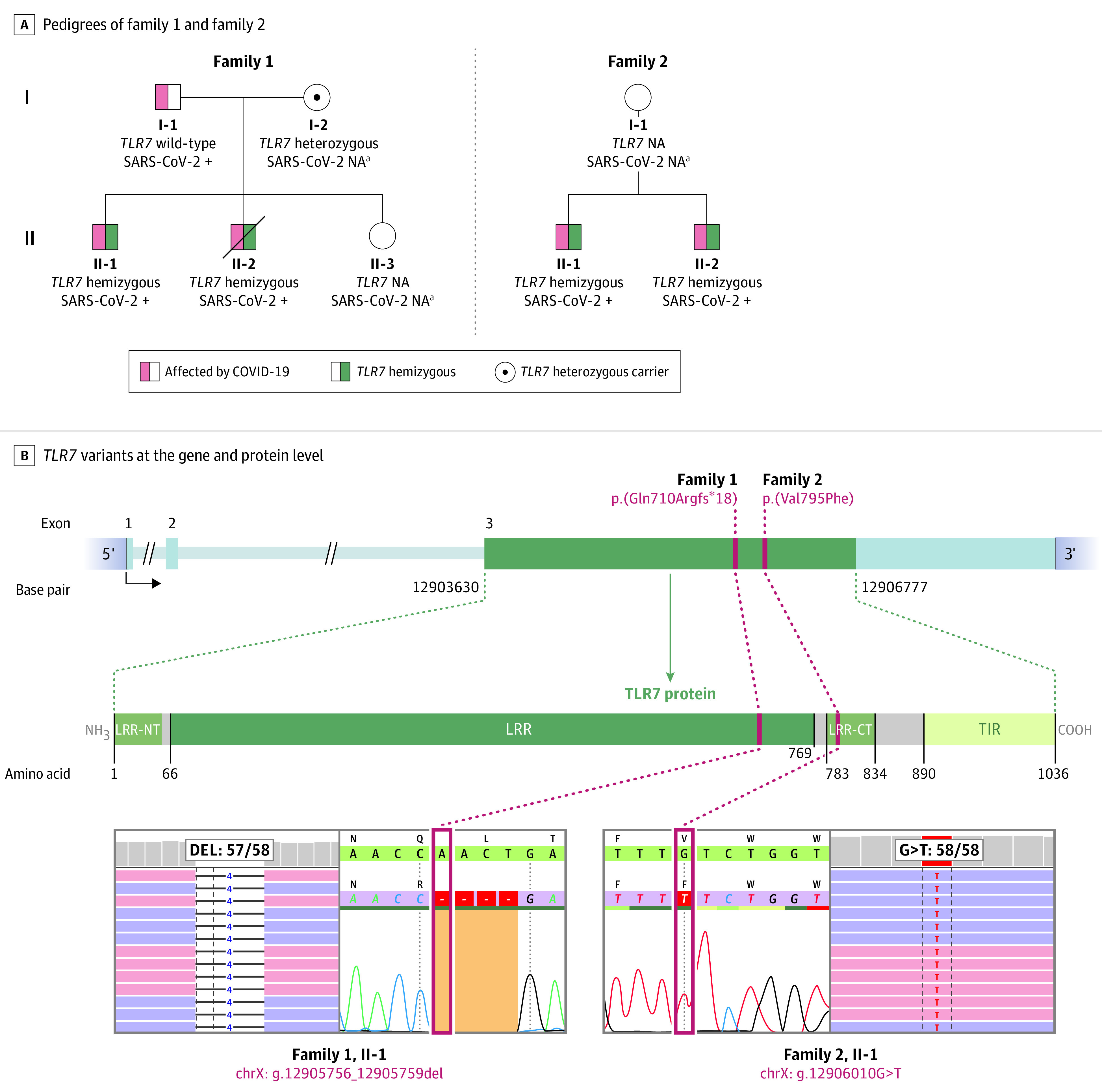
Design, setting, and participants: Case series of pairs of brothers without medical history meeting the selection criteria of young (age <35 years) brother pairs admitted to the intensive care unit (ICU) due to severe COVID-19. Four men from 2 unrelated families were admitted to the ICUs of 4 hospitals in the Netherlands between March 23 and April 12, 2020. The final date of follow-up was May 16, 2020. Available family members were included for genetic variant segregation analysis and as controls for functional experiments.

Exposure: Severe COVID-19.

Main outcome and measures: Results of rapid clinical whole-exome sequencing, performed to identify a potential monogenic cause. Subsequently, basic genetic and immunological tests were performed in primary immune cells isolated from the patients and family members to characterize any immune defects.

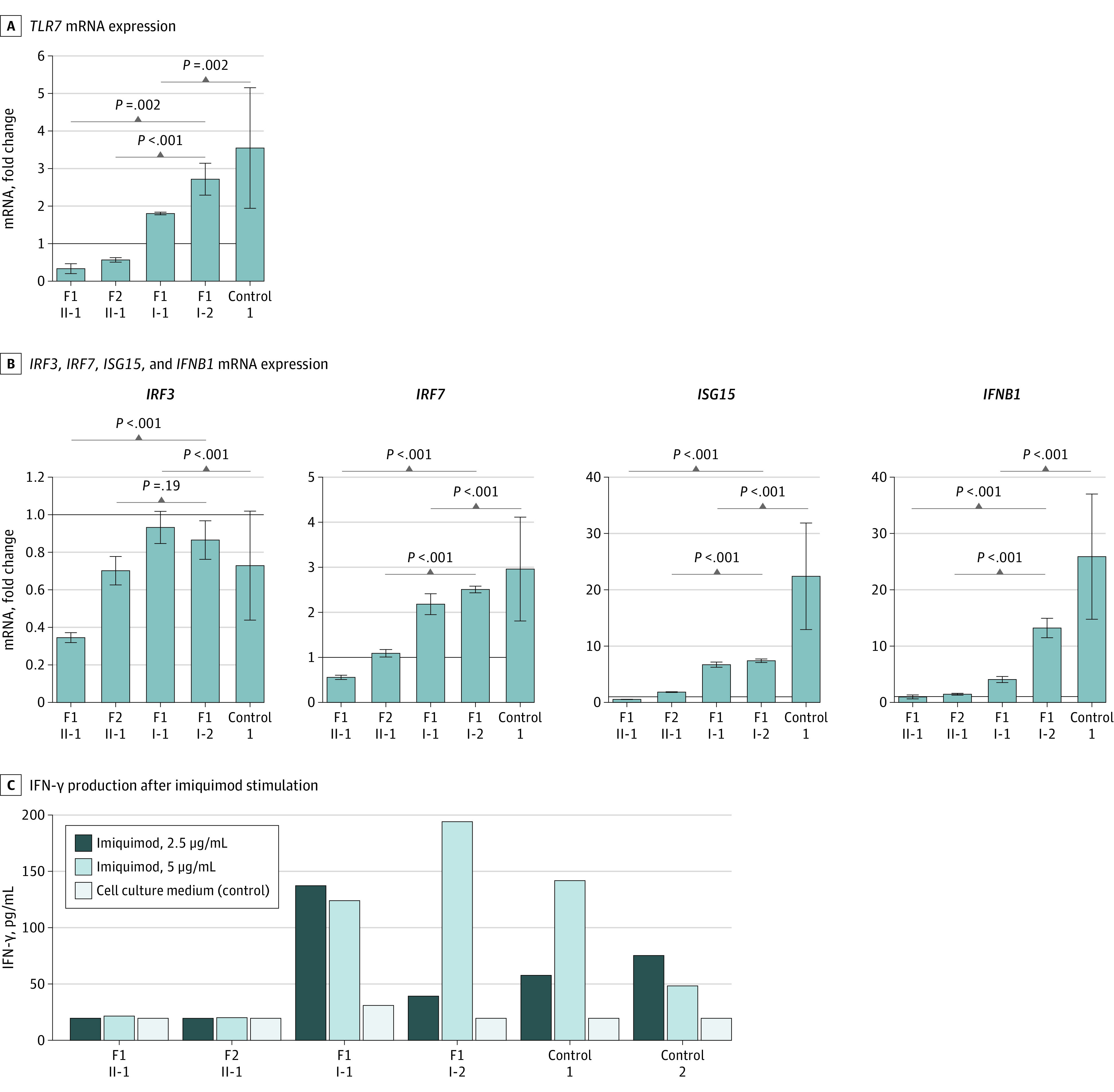
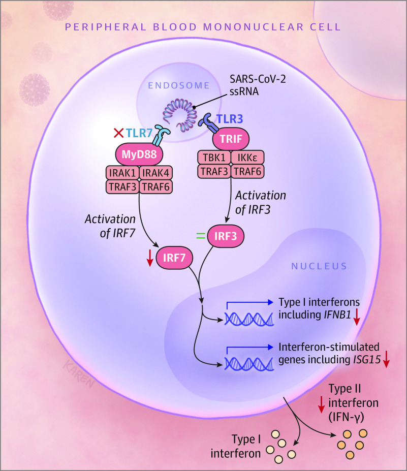
Results: The 4 male patients had a mean age of 26 years (range, 21-32), with no history of major chronic disease. They were previously well before developing respiratory insufficiency due to severe COVID-19, requiring mechanical ventilation in the ICU. The mean duration of ventilatory support was 10 days (range, 9-11); the mean duration of ICU stay was 13 days (range, 10-16). One patient died. Rapid clinical whole-exome sequencing of the patients and segregation in available family members identified loss-of-function variants of the X-chromosomal TLR7. In members of family 1, a maternally inherited 4-nucleotide deletion was identified (c.2129_2132del; p.[Gln710Argfs*18]); the affected members of family 2 carried a missense variant (c.2383G>T; p.[Val795Phe]). In primary peripheral blood mononuclear cells from the patients, downstream type I interferon (IFN) signaling was transcriptionally downregulated, as measured by significantly decreased mRNA expression of IRF7, IFNB1, and ISG15 on stimulation with the TLR7 agonist imiquimod as compared with family members and controls. The production of IFN-γ, a type II IFN, was decreased in patients in response to stimulation with imiquimod.

Conclusions and relevance: In this case series of 4 young male patients with severe COVID-19, rare putative loss-of-function variants of X-chromosomal TLR7 were identified that were associated with impaired type I and II IFN responses. These preliminary findings provide insights into the pathogenesis of COVID-19.


Conflict of interest statement

Conflict of Interest Disclosures: Dr Joosten reported being a scientific founder of Trained Therapeutic Discovery and a scientific advisory board member of Olatec Therapeutics. Dr Netea reported being a scientific founder of Trained Therapeutic Discovery and receiving grants from ViiV HealthCare outside the submitted work. No other disclosures were reported.

Figures

Figure 1.
Figure 1.. Identification of TLR7 Variants in 4 Patients From 2 Families With Severe Coronavirus Disease 2019 (COVID-19)
Panel A shows the pedigrees of the 2 families and respective segregation of TLR7 variants as well as the COVID-19 status if determined. Patients II-1 and II-2 from family 1 lived in separate households, patients II-1 and II-2 from family 2 were housed together. Circles represent female family members; squares, males. A slash symbol represents a deceased individual. Panel B shows the TLR7 variants in each family at the gene and protein level in a schematic representation. The TLR7 protein structure is shown with leucine-rich repeat region (LRR), N- and C-termini (LRR-NT, LRR-CT) and the toll-interleukin receptor (TIR) homology domain. The exon-intron structure depicts the coding exon 3 of TLR7 with the identified variants by exome sequencing and Sanger sequencing validation as shown in the highlighted sections below. Red boxes depict positions of the variants ChrX(GRCh37):g.12905756_12905759del and ChrX(GRCh37):g.12906010G>T). NA indicates not assessed. aAt the time of evaluation, testing for severe acute respiratory syndrome coronavirus 2 (SARS-CoV-2) was not routinely performed.
Figure 2.
Figure 2.. Assessment of Type I and II Interferon (IFN) Responses in Peripheral Blood Mononuclear Cells Derived From Patients and Controls
Panel A shows TLR7 mRNA expression in both index patients compared with the parents of family 1 and healthy controls after imiquimod stimulation for 4 hours (5 μg/mL) in comparison with negative controls (cell culture medium, Roswell Park Memorial Institute [RPMI]). Panel B shows the fold change in mRNA expression of type I IFN–related genes IRF3, IRF7, ISG15, and IFNB1 induced by TLR7 agonist imiquimod (5 μg/mL) as compared with negative controls. The solid lines in panels A and B signify a fold change of 1. Panel C shows the production of IFN-γ production after imiquimod stimulation for 7 days in concentrations of 2.5 μg/mL and 5 μg/mL as compared with unstimulated control cells (RPMI). Results depicted in panels A and B have been replicated in an independent experiment for the brother of II-2 from family 2; eFigure 4 in the Supplement). Control 1 in panels A, B, and C represents a healthy female; control 2 in panel C refers to a healthy male. Error bars show standard deviations. P values were calculated using a t test performed on log-transformed fold changes. F1 indicates family 1; F2, family 2; IRF3, interferon regulatory factor 3; IRF7, interferon regulatory factor 7; ISG15, interferon-stimulated gene 15; and IFNB1, interferon beta 1.
Figure 3.
Figure 3.. Illustration of a Proposed Role for Impaired TLR7 Signaling Induced by Loss-of-Function Variants in Peripheral Blood Mononuclear Cells
TLR7 signaling is induced by endocytosed severe acute respiratory syndrome coronavirus 2 (SARS-CoV-2) single-stranded RNA (ssRNA) motifs. Downstream effects are transduced by the adapter MyD88, associating kinases IRAK1 and IRAK4, and ubiquitin ligases TRAF3 and TRAF6. This is followed by activation of interferon regulatory factor 7 (encoded by IRF7, OMIM 605047), which translocates to the nucleus to induce transcription of type I interferon (including interferon beta 1 [IFNB1, OMIM 147640] and interferon-stimulated genes [ISGs], including ISG15 [OMIM 147571]). In parallel, the endosomal TLR3 receptor senses SARS-CoV-2 ssRNA and consequently activates adapter protein TRIF, which then recruits associating kinases TBK1 and IKKε and the ubiquitin ligases TRAF3 and TRAF6. TLR3 signaling continues via stimulation of IFN regulatory factor 3 (encoded by IRF3, OMIM 300365), which transfers to the nucleus to induce transcription of type I IFN genes. In TLR7 deficiency due to loss-of-function variants (as indicated by the red cross), transcription of downstream effector genes IRF7, IFNB1, and ISG15 are downregulated (as indicated by downward pointing arrows). In addition, the production of IFN-γ is impaired. The expression of IRF3 remains unaltered (as indicated by the equal sign).

Comment in

References

    1. Klompas M, Baker MA, Rhee C. Airborne transmission of SARS-CoV-2: theoretical considerations and available evidence. JAMA. 2020. doi:10.1001/jama.2020.12458 - DOI - PubMed
    1. Richardson S, Hirsch JS, Narasimhan M, et al. ; and the Northwell COVID-19 Research Consortium . Presenting characteristics, comorbidities, and outcomes among 5700 patients hospitalized with COVID-19 in the New York City area. JAMA. 2020;323(20):2052-2059. doi:10.1001/jama.2020.6775 - DOI - PMC - PubMed
    1. Grasselli G, Zangrillo A, Zanella A, et al. ; COVID-19 Lombardy ICU Network . Baseline characteristics and outcomes of 1591 patients infected with SARS-CoV-2 admitted to ICUs of the Lombardy Region, Italy. JAMA. 2020;323(16):1574-1581. doi:10.1001/jama.2020.5394 - DOI - PMC - PubMed
    1. Guan WJ, Ni ZY, Hu Y, et al. ; China Medical Treatment Expert Group for Covid-19 . Clinical characteristics of coronavirus disease 2019 in China. N Engl J Med. 2020;382(18):1708-1720. doi:10.1056/NEJMoa2002032 - DOI - PMC - PubMed
    1. Tay MZ, Poh CM, Rénia L, MacAry PA, Ng LFP. The trinity of COVID-19: immunity, inflammation and intervention. Nat Rev Immunol. 2020;20(6):363-374. doi:10.1038/s41577-020-0311-8 - DOI - PMC - PubMed

Publication types