Review
. 2021 Jan 15;22(2):288-297.
doi: 10.1002/cbic.202000376. Epub 2020 Aug 28.

On the Case of the Misplaced Hydrogens

Affiliations
Review

On the Case of the Misplaced Hydrogens

Prashasti Kumar et al. Chembiochem. .

Abstract

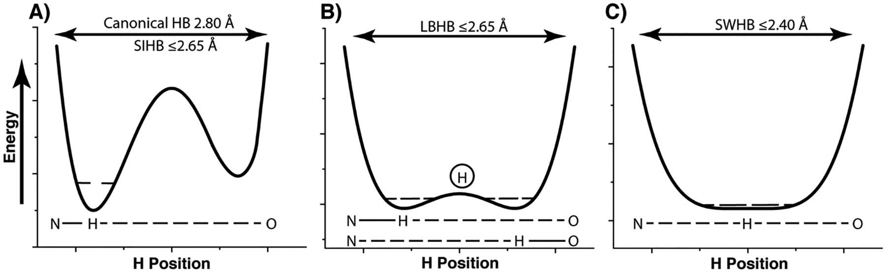
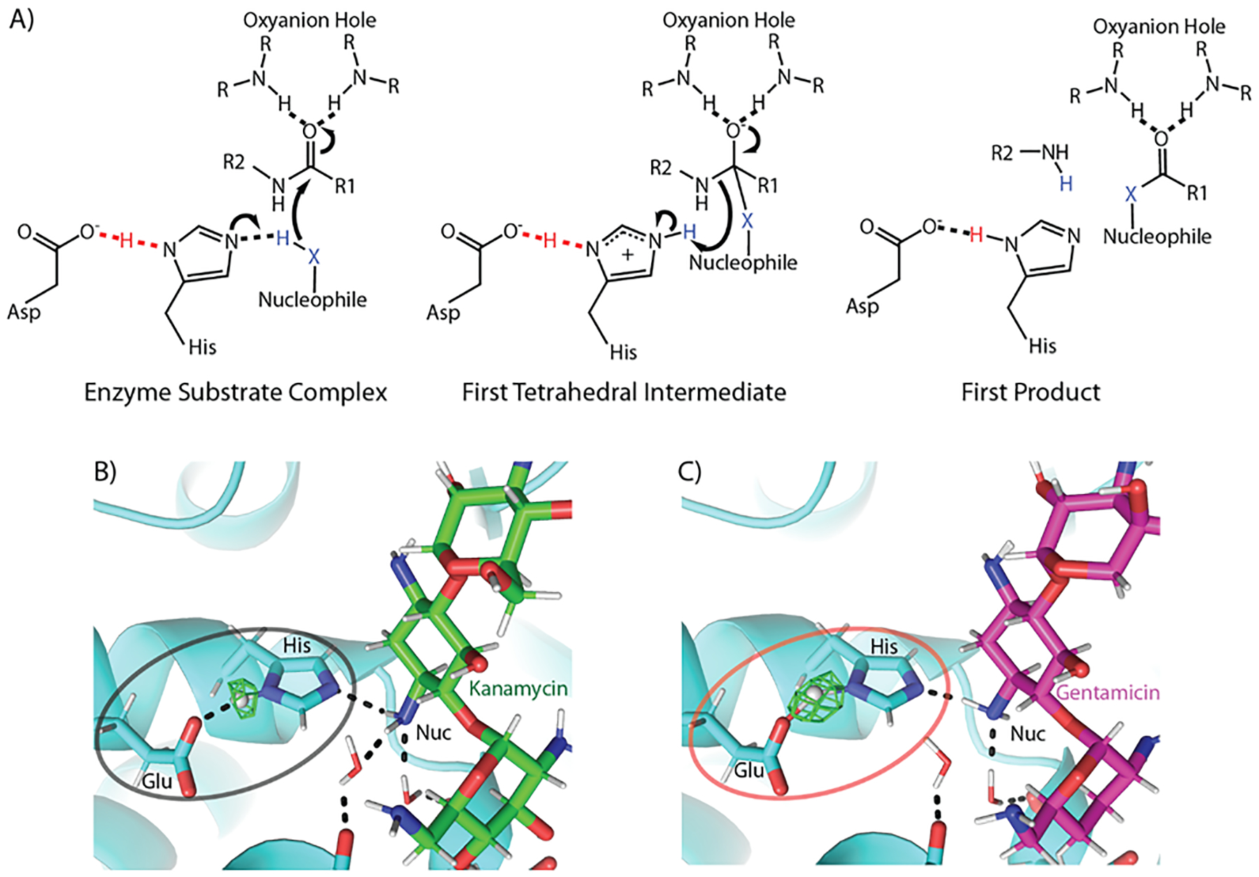
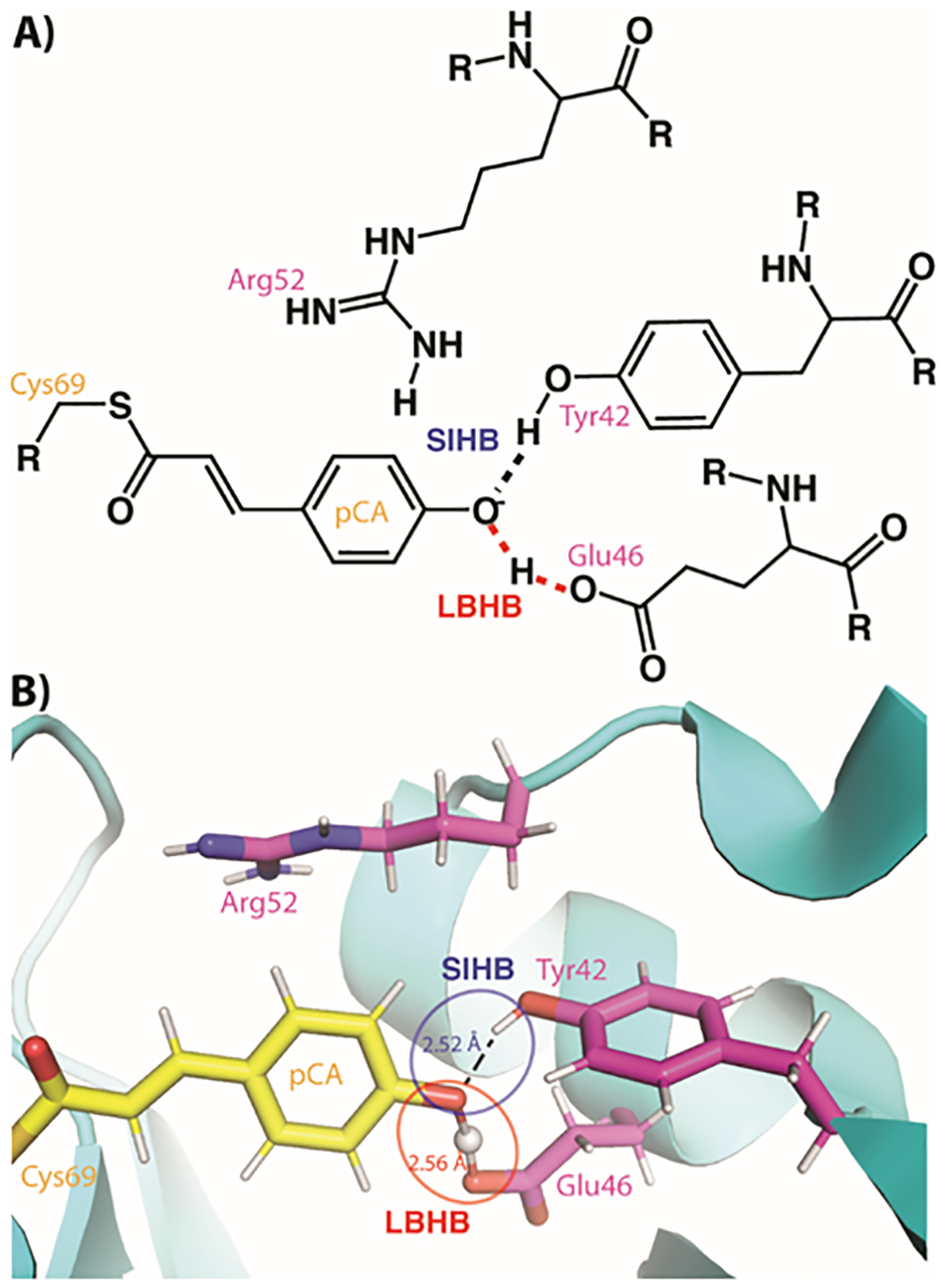
Few other elements play a more central role in biology than hydrogen. The interactions, bonding and movement of hydrogen atoms are central to biological catalysis, structure and function. Yet owing to the elusive nature of a single hydrogen atom few experimental and computational techniques can precisely determine its location. This is exemplified in short hydrogen bonds (SHBs) where the location of the hydrogen atom is indicative of the underlying strength of the bonds, which can vary from 1-5 kcal/mol in canonical hydrogen bonds, to an almost covalent nature in single-well hydrogen bonds. Owing to the often-times inferred position of hydrogen, the role of SHBs in biology has remained highly contested and debated. This has also led to discrepancies in computational, biochemical and structural studies of proteins thought to use SHBs in performing chemistry and stabilizing interactions. Herein, we discuss in detail two distinct examples, namely the conserved catalytic triad and the photoreceptor, photoactive yellow protein, where studies of these SHB-containing systems have permitted contextualization of the role these unique hydrogen bonds play in biology.

Keywords: catalytic triads; hydrogen bonds; low-barrier hydrogen bonds; photoactive yellow proteins; short ionic hydrogen bonds.


Figures

Figure 1.
Figure 1.. Schematic of typical potential energy profiles for different types of hydrogen bonds.
A) In a canonical hydrogen bond, an asymmetric double well potential exists, where the hydrogen remains attached to the donor heavy atom. In a short-ionic hydrogen bond (SIHB), the asymmetric double well persists even though the distance between the atoms can be less than the van der Waals radii. B) In a low barrier hydrogen bond (LBHB) matching pKa values of the donor and acceptor heavy atoms means that the transfer of the hydrogen to either heavy atoms occurs with an equal probability and the average position of the hydrogen atom is in the center, as seen in structural studies. C) In single-well hydrogen bond (SWHB) the energetic barrier for transfer of the hydrogen atom is lost completely and it is simultaneously bound to both the heavy atoms. The dashed lines within the wells represent the zero-point energies.
Figure 2.
Figure 2.. Low barrier hydrogen bonds in catalytic triads.
A) Three states of a general catalytic triad mechanism are shown. In the starting enzyme substrate complex, a hydrogen bond exists between the aspartic acid and the histidine Nδ, an interaction found in some instances to be an LBHB, increases the basicity of the histidine Nε (left). This permits the abstraction of a proton from the nucleophile, which subsequently attacks the peptide bond and generates the first tetrahedral intermediate (center). The first intermediate collapses resulting in the first two products of the reaction (right). R groups indicate continuation of the protein chain. B) The local chemical environment of the non-canonical (Glu-His-antibiotic amine) catalytic triad found in an aminoglycoside acetyltransferase dictates the type of hydrogen bond when bound to B) kanamycin and C) gentamicin. The Fo-Fc nuclear omit density for the hydrogen atom involved in the hydrogen bond between the catalytic residues is shown in green. For the least catalytically preferred substrate (kanamycin), a canonical hydrogen bond is found, whereas for one of the best turned over antibiotics (gentamicin) a low barrier hydrogen bond is found.
Figure 3.
Figure 3.. The chromophore binding site in photoactive yellow protein (PYP).
A) A schematic of the PYP chromophore (pCA) binding site with the two types of SHBs indicated. R groups indicate continuation of the protein chain. B) The neutron crystal structure of the PYP chromophore active site with the two types of SHBs indicated.

References

    1. Herschlag D, Pinney MM, Biochemistry 2018, 57, 3338–3352. - PubMed
    1. Frey PA, Whitt SA, Tobin JB, Science 1994, 264, 1927–1930; - PubMed
    2. Cleland WW, Frey PA, Gerlt JA, Journal of Biological Chemistry 1998, 273, 25529–25532. - PubMed
    1. Cleland WW, Arch Biochem Biophys 2000, 382, 1–5. - PubMed
    1. Kreevoy MM, Liang TM, Journal of the American Chemical Society 1980, 102, 3315–3322;
    2. Cleland WW, Biochemistry 1992, 31, 317–319; - PubMed
    3. Gerlt JA, Kreevoy MM, Cleland W, Frey PA, Chem Biol 1997, 4, 259–267. - PubMed
    1. Gerlt JA, Gassman PG, Journal of the American Chemical Society 1993, 115, 11552–11568;
    2. Gerlt JA, Gassman PG, Biochemistry 1993, 32, 11943–11952; - PubMed
    3. Cleland WW, Kreevoy MM, Science 1994, 264, 1887–1890. - PubMed

Publication types

LinkOut - more resources