. 2020 Aug 11;15(2):482-497.
doi: 10.1016/j.stemcr.2020.06.019. Epub 2020 Jul 23.

Imaging Transplanted Photoreceptors in Living Nonhuman Primates with Single-Cell Resolution

Affiliations

Imaging Transplanted Photoreceptors in Living Nonhuman Primates with Single-Cell Resolution

Ebrahim Aboualizadeh et al. Stem Cell Reports. .

Abstract

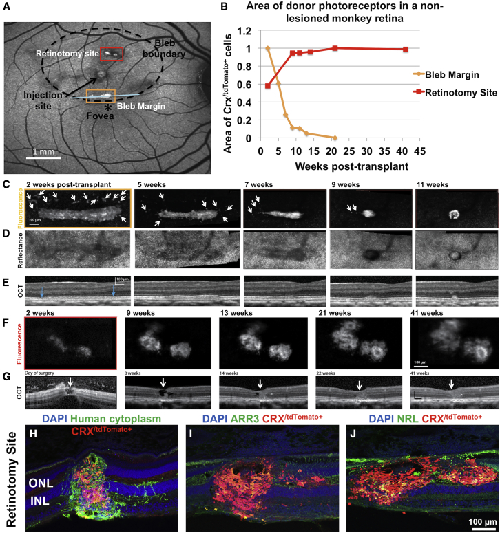
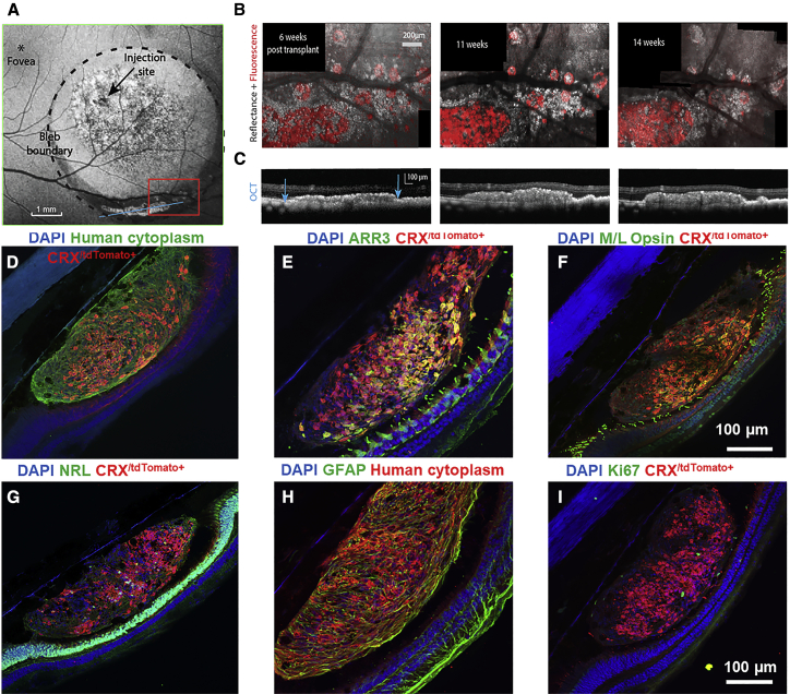
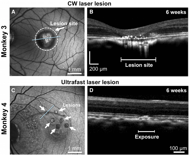
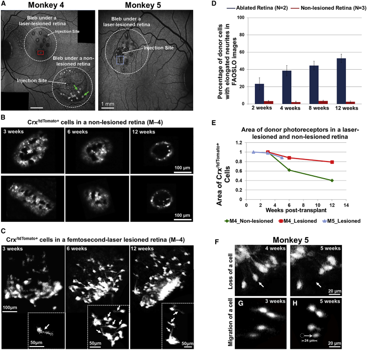
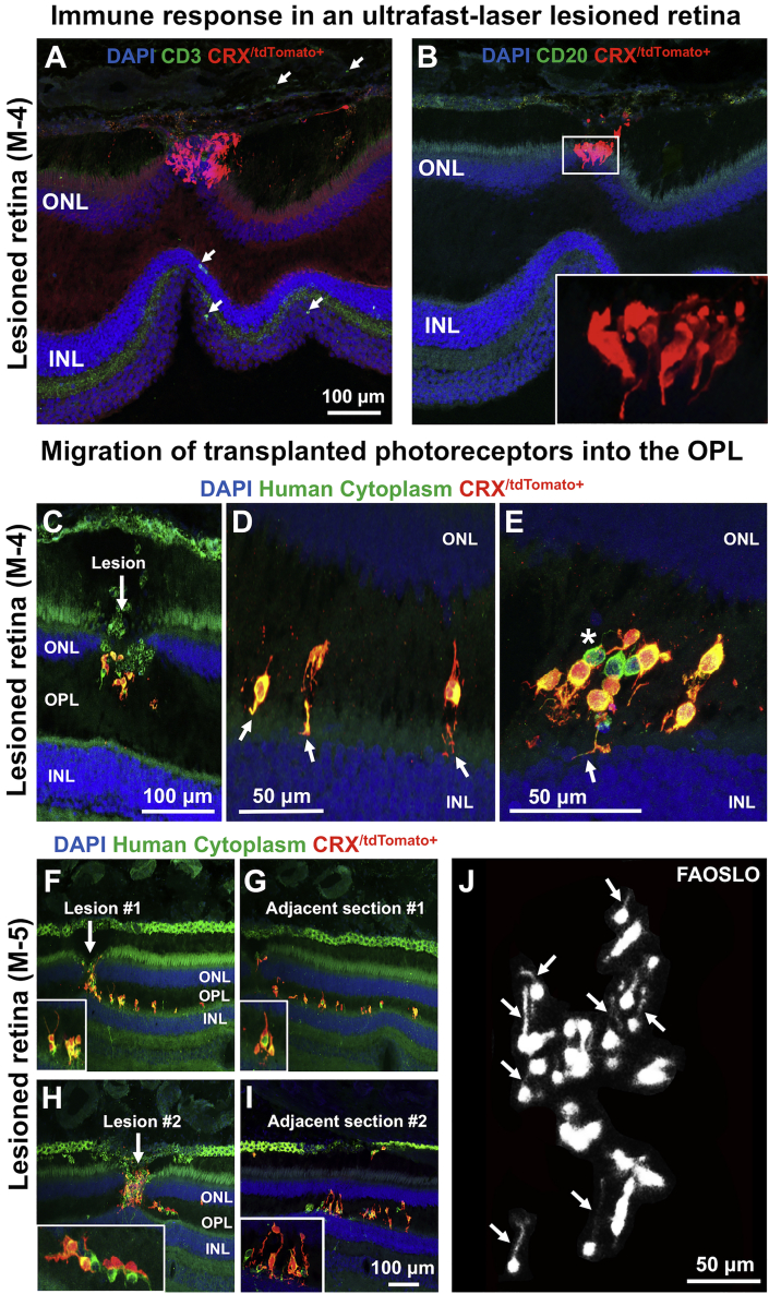
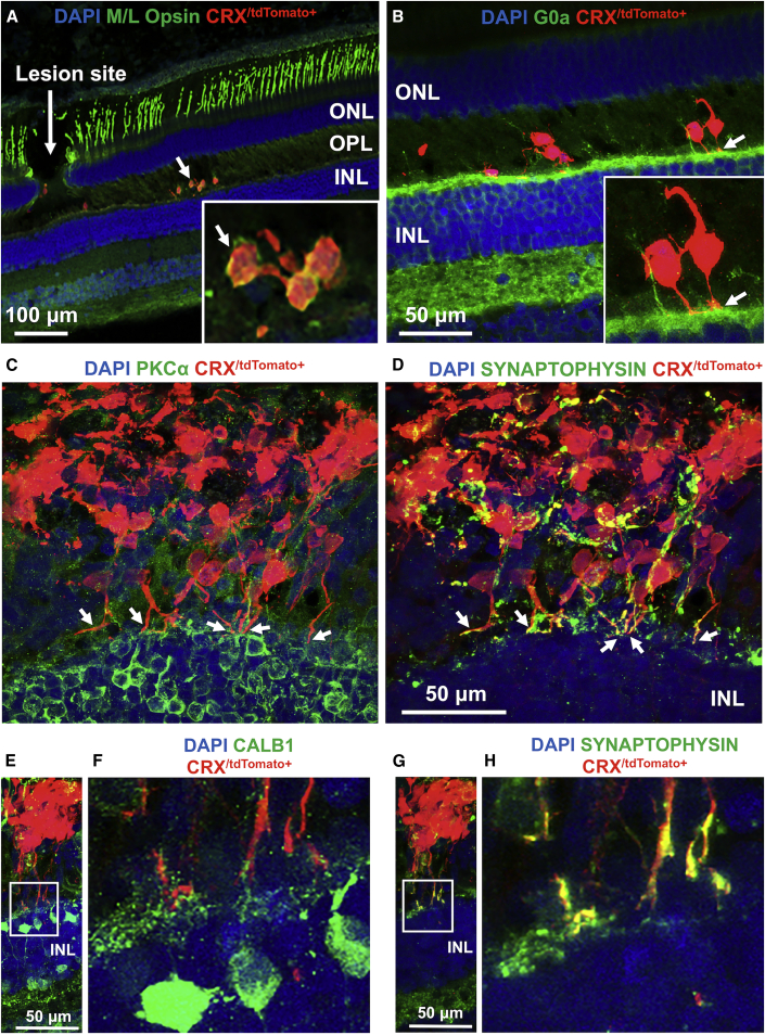
Stem cell-based transplantation therapies offer hope for currently untreatable retinal degenerations; however, preclinical progress has been largely confined to rodent models. Here, we describe an experimental platform for accelerating photoreceptor replacement therapy in the nonhuman primate, which has a visual system much more similar to the human. We deployed fluorescence adaptive optics scanning light ophthalmoscopy (FAOSLO) to noninvasively track transplanted photoreceptor precursors over time at cellular resolution in the living macaque. Fluorescently labeled photoreceptors generated from a CRX+/tdTomato human embryonic stem cell (hESC) reporter line were delivered subretinally to macaques with normal retinas and following selective ablation of host photoreceptors using an ultrafast laser. The fluorescent reporter together with FAOSLO allowed transplanted photoreceptor precursor survival, migration, and neurite formation to be monitored over time in vivo. Histological examination suggested migration of photoreceptor precursors to the outer plexiform layer and potential synapse formation in ablated areas in the macaque eye.

Keywords: adaptive optics retinal imaging; fluorescence; hPSC; integration and survival; in vivo; nonhuman primates; photoreceptor precursor; retinal degeneration; stem cell therapy; ultrafast.


Figures

Figure 1
Figure 1
Long-Term Evaluation of Transplanted Photoreceptor Precursors in a Normal, Non-lesioned Monkey Retina (A) Following transplantation, SLO imaging of the retina 4 weeks post transplant shows three regions with fluorescent cells, two at or near retinotomies associated with injection attempts, and one at the inferior aspect of the bleb. Bleb boundary (dotted circle) and injection site (black arrow) are shown. Retinotomy site refers to the incision created during failed attempts to raise a bleb. (B) Quantification of two areas of transplanted photoreceptors using FAOSLO. There is a gradual loss of individual cells in the main cell cluster at the inferior margin of the bleb; however, at the retinotomy site there was an increase in the the area of the cell clusterfrom 2 to 9 weeks, followed by apparent stabilization up to 41 weeks post transplantation. (C) FAOSLO imaging tracking a tdTomato labelled cell cluster at the inferior mergin of the bleb (yellow box in A) over an 11-week period. White arrows show loss of single cells and small clusters over time. (D) NIR reflectance AOSLO imaging showed the photoreceptor mosaic in the same region as (C). (E) OCT showed the axial changes in the transplant shown in (C) over time with blue arrows indicating the location of the cell cluster. (F) FAOSLO images tracking a tdTomato labelled cell cluster at the retinotomy site (red box in A) over a 41-week period are shown. (G) OCT showing axial changes in the transplant shown in (F) at the retinotomy site a 41 week period. White arrows show the hole that was created at the retinotomy site. As shown, significant cell efflux was observed, which we attribute to raising the bleb and injecting donor cells simultaneously in this case. (H–J) Immunohistochemistry at 41 weeks post transplant demonstrated the presence of transplanted cells (human cytoplasm+), including CRX/tdTomato+ photoreceptors, which filled the retinotomy site. (I and J) Transplanted photoreceptors matured in vivo to express cone (I) and rod (J) markers.
Figure 2
Figure 2
Photoreceptor Precursors in a Non-lesioned Retina Remained Confined to the Subretinal Space (A) A blue autofluorescence SLO image of the bleb and transplanted cells is shown. Autofluorescence from disrupted RPE can be seen in the central bleb as well as tdTomato fluorescence from the transplanted cells at the inferior margin of the bleb. The region bounded by the red box was imaged using FAOSLO and AOSLO NIR reflectance imaging for 14 weeks. (B) Merged fluorescence (red) and NIR reflectance (gray) AOSLO images taken at 6, 11, and 14 weeks post transplant are shown. (C) OCT of the region containing the transplanted cells along an axis corresponding to the blue line shown in (A) is shown. (D–I) CRX/tdTomato+ cells always co-labeled with a human cytoplasm-specific antibody, confirming that these cells were of human origin. Additional retinal cell types from the dissociated organoids were also present (i.e., human cytoplasm+/CRX/tdTomato-cells). Photoreceptor precursors demonstrated expression of cone (E and F) and rod markers (G), Some human cytoplasm+/GFAP+ glial cells were also present within the cell cluster (H), while proliferative cells were minimal (I).
Figure 3
Figure 3
Evaluation of the Retinal Impact of Exposure to a CW Laser and an Ultrafast Laser Using SLO and OCT (A and B) Conventional SLO (A) and a spectral-domain OCT scan (B) of a monkey retina ablated with a CW laser at 6 weeks post exposure. At the lesion site, there is complete ablation of the outer retina with apposition of the inner plexiform layer to the RPE. The RPE is irregular with overlying scattered hyperreflective foci. OCT scan was acquired in the area designated by the blue line in (A). (C and D) SLO (C) and a spectral-domain OCT scan (D) of a monkey retina ablated using an ultrafast laser with adaptive optics at 6 weeks post exposure. OCT imaging shows localized disruption within the outer limiting membrane and the outer nuclear layer. The linear dimension of an exposure area is shown on the OCT scan. OCT was acquired across the blue line in (C). White arrows in (C) highlight the pattern of ultrafast laser lesions.
Figure 4
Figure 4
FAOSLO Imaging of Photoreceptor Precursors in the AO Ultrafast Nonhuman Primate Model (A) SLO images of photoreceptor precursors in monkeys 4 and 5, 3 weeks post transplantation are shown. Monkey 4 was tracked with in vivo FAOSLO imaging for 12 weeks and monkey 5 was tracked for 5 weeks. For a direct comparison within the same eye in monkey 4, one transplant was performed in the lesioned area and one was performed in a non-lesioned area. Monkey 5 received a transplant into a lesioned area. (B and C) FAOSLO images of transplanted cells in a non-lesioned retina (B) and an AO ultrafast-laser-lesioned retina are shown (C) at 3, 6, and 12 weeks post transplant. (B) FAOSLO imaging of two cell clusters in a non-lesioned retina (green arrows in A) showed loss of donor cells over time. (C) By comparison, transplanted cells in the lesioned area extended neuronal processes over time are shown (see also Figure S1). Fine neurites could be resolved on individual photoreceptor precursors in this region with FAOSLO (white arrows). (D) Area of transplanted cells in two monkeys and both blebs were quantified, showing pattern of cell loss in both ablated and non-lesioned retinas. (E) The percentage of single cells with extended neurites in the lesioned retinas (N = 2) varied between 22% and 52%; however, in non-lesioned retinas (N = 3), this percentage was less than 8% over a 12-week period. (F–H) (F) Single-cell tracking using FAOSLO imaging showed loss of a single photoreceptor precursor between 4 and 5 weeks post transplant (white arrows) and migration of a different photoreceptor precursor over a 2-week period (G). White dotted circle in (H) shows the original position of a single cell at 3 weeks and the white arrow shows the direction of migration. Errors bars represent standard error and N represents number of animals.
Figure 5
Figure 5
Immunohistochemical Examination of Photoreceptor Precursors in the AO Ultrafast-Lesioned Retina (A and B) Histological examination of transplanted cells in an ultrafast-lesioned retina in monkey 4 is shown. In this model, very few anti CD3 T cells (A) and anti CD20 B cells (B) were noted (see also Figure S2). Transplanted cells in higher magnification are shown in the inset. (C) In monkey 4, histology at 12 weeks showed that transplanted photoreceptors had migrated into the OPL through the laser lesion site. (D) Some donor cells extended neurites toward the INL through the entry site provided by laser lesions (white arrows). (E) Additional cells that had migrated to the OPL and contacted the host INL by extending neurites are shown (White arrow). Asterisk in (E) shows human non-photoreceptors. (F) In monkey 5, similar migration of transplanted photoreceptor precursors into the host OPL was noted at the ablation site within a shorter period of time (5 weeks, cells are shown at higher magnification in the insets). (G) The Adjacent section also showed transplanted cells that migrated to the OPL through the laser lesions and extended neuronal processes. (H) Migration of donor photoreceptors into the OPL through the second lesion site is shown. (I) Similarly, the adjacent section showed migration of donor cells to the OPL and extension of neuronal processes (higher magnification images are shown in the insets). (J) FAOSLO imaging at the same retinal location, 1 week before euthanasia, showing the capability of FAOSLO to resolve fine neurites extending laterally in vivo.
Figure 6
Figure 6
Immunohistochemical Examination of Photoreceptor Precursor Integration and Differentiation (A) Some migratory transplanted photoreceptor precursors matured in vivo to express M/L opsin. (B) Neurites from transplanted cells were in contact with host second-order neurons, including G0α+ ON bipolar cell dendrites. (C and D) Some donor photoreceptor precursors extended axons toward host bipolar cells (PKCα+, C) and expressed the presynaptic protein marker (synaptophysin) (D). While expression of synaptophysin in transplanted cells was disorganized in cells away from the host OPL, processes close to the host INL had pronounced expression of synaptophysin in putative axonal terminals. (E–H) Some photoreceptor precursors contacted host second-order neurons, including CALB1+ horizontal and bipolar cells (E and F) and expressed synaptophysin (G and H). The white squares in (E) and (G) are shown magnified in (F) and (H), respectively.

References

    1. Barber A.C., Hippert C., Duran Y., West E.L., Bainbridge J.W., Warre-Cornish K., Luhmann U.F., Lakowski J., Sowden J.C., Ali R.R., Pearson R.A. Repair of the degenerate retina by photoreceptor transplantation. Proc. Natl. Acad. Sci. U S A. 2013;110:354–359. - PMC - PubMed
    1. Busskamp V., Picaud S., Sahel J.A., Roska B. Optogenetic therapy for retinitis pigmentosa. Gene Ther. 2012;19:169. - PubMed
    1. Chao J.R., Lamba D.A., Klesert T.R., La Torre A., Hoshino A., Taylor R.J., Jayabalu A., Engel A.L., Khuu T.H., Wang R.K., Neitz M. Transplantation of human embryonic stem cell-derived retinal cells into the subretinal space of a non-human primate. Transl. Vis. Sci. Technol. 2017;6:4. - PMC - PubMed
    1. da Cruz L., Fynes K., Georgiadis O., Kerby J., Luo Y.H., Ahmado A., Vernon A., Daniels J.T., Nommiste B., Hasan S.M., Gooljar S.B. Phase 1 clinical study of an embryonic stem cell-derived retinal pigment epithelium patch in age-related macular degeneration. Nat. Biotechnol. 2018;36:328. - PubMed
    1. Dhakal K.R., Walters S., McGregor J.E., Schwarz C., Strazzeri J.M., Aboualizadeh E., Bateman B., Huxlin K.R., Hunter J.J., Williams D.R., Merigan W.H. Localized photoreceptor ablation using femtosecond pulses focused with adaptive optics. Transl. Vis. Sci. Technol. 2020;9:16. - PMC - PubMed

Publication types

LinkOut - more resources