Review
. 2020:157:37-62.
doi: 10.1016/j.addr.2020.07.012. Epub 2020 Jul 22.

Micro and nanoscale technologies in oral drug delivery

Affiliations
Review

Micro and nanoscale technologies in oral drug delivery

Samad Ahadian et al. Adv Drug Deliv Rev. 2020.

Abstract

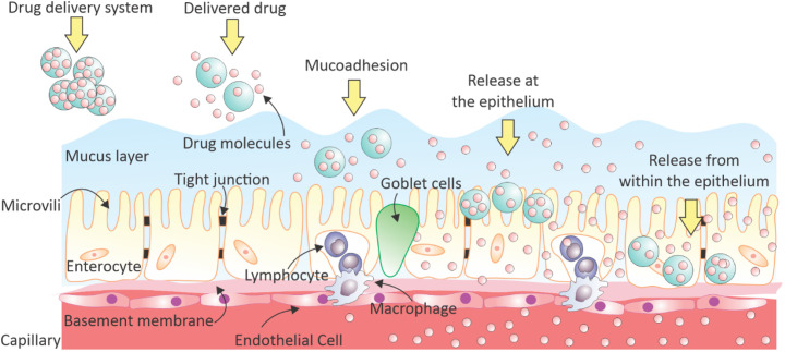
Oral administration is a pillar of the pharmaceutical industry and yet it remains challenging to administer hydrophilic therapeutics by the oral route. Smart and controlled oral drug delivery could bypass the physiological barriers that limit the oral delivery of these therapeutics. Micro- and nanoscale technologies, with an unprecedented ability to create, control, and measure micro- or nanoenvironments, have found tremendous applications in biology and medicine. In particular, significant advances have been made in using these technologies for oral drug delivery. In this review, we briefly describe biological barriers to oral drug delivery and micro and nanoscale fabrication technologies. Micro and nanoscale drug carriers fabricated using these technologies, including bioadhesives, microparticles, micropatches, and nanoparticles, are described. Other applications of micro and nanoscale technologies are discussed, including fabrication of devices and tissue engineering models to precisely control or assess oral drug delivery in vivo and in vitro, respectively. Strategies to advance translation of micro and nanotechnologies into clinical trials for oral drug delivery are mentioned. Finally, challenges and future prospects on further integration of micro and nanoscale technologies with oral drug delivery systems are highlighted.

Keywords: Drug delivery devices; Micro and nanocarriers; Micro and nanoscale technologies; Oral drug delivery; Tissue models.


Figures

Unlabelled Image
Graphical abstract
Fig. 1
Fig. 1
Micro- and nanoscale technologies enable fabrication of oral drug carriers as well as human tissue-on-a-chip models for precision medicine applications.
Fig. 2
Fig. 2
Schematic illustration of drug release and absorption mechanisms for orally delivered drugs in the large surface area of human intestinal epithelium.
Fig. 3
Fig. 3
A schematic of physiological barriers in oral drug delivery including: (a) biochemical barriers, (b) mucus barrier, and (c) cellular barriers to oral drug delivery. Reprinted by permission from Springer Nature [26] Copyright (2019).
Fig. 4
Fig. 4
Microfluidic approaches to fabricate nanocarriers for oral drug delivery. Different diffusion- and droplet-based microfluidic platforms for preparation of nanoparticles including (a) microfluidic continuous flow, (b) microfluidic mixer, (c) microfluidic droplet generator, (d) microfluidic processor. Reprinted from [57] Copyright (2013), with permission from Elsevier.
Fig. 5
Fig. 5
Fabrication and characterization of nanocarriers for oral drug delivery. (a) Schematic image of possibilities for drug loading and functionalization with different targeting and therapeutics ligands in liposomes. Reprinted from [92] with permission from Elsevier. (b) A strategy for loading hydrophilic drugs in the core of solid nanoparticles (blue color) by generation of a hydrophilic viscose phase in the core. Reprinted from [101], Copyright (2016), with permission from Elsevier. (c) A two-step preparation method for insulin-loaded core-shell nanoparticles composed of a modified chitosan core coated with thiolated hyaluronic acid through electrostatic [114]. Copyright (2018) Wiley-VCH Verlag GmbH & Co. KGaA. Reproduced with permission. (d) Self-assembly of cationic copolymers (yellow color) with anion biomacromolecules (green color) to form polymer micelles with targeting agents can improve mucoadhesion and can generate polymeric networks of micelles. Reprinted (adapted) with permission from [115] Copyright (2005) American Chemical Society.
Fig. 6
Fig. 6
Micropatches in oral drug delivery. (a) Schematic representation showing a GI patch and (b) working mechanism of the hard capsule filled with mucoadhesive patches. Reprinted from [152], Copyright (2011), with permission from Elsevier. (c) In contrast to microspheres, asymmetric and planar microdevices facilitate proximal and unidirectional drug release, while increasing residence time in the GI tract. Reprinted from [29], Copyright (2015), with permission from Elsevier.
Fig. 7
Fig. 7
Examples of fabricated devices for oral drug delivery. (a) Caplets fabricated by 3D printing with various designs of multiple materials showed by different colors. Reprinted with permission from [160]. Copyright (2016) American Chemical Society. (b) 3D printed multi-compartment capsular devices with two phase release profiles. Reprinted from [158] Copyright (2017), with permission from Elsevier. (c) The device consisted of an elastomeric part (core) and six drug-loaded arms. Various polymers (blue, red and yellow) released the drug at different rates. Material from [161], published 2018, Nature Springer. (d) Scanning electron microscopic (SEM) image of the microcontainer filled with polymer and impregnated with ketoprofen. Scale bar is 100μm. Reprinted from [163] Copyright (2014), with permission from Elsevier. (e) Schematic of the drug- loaded micromotor and drug delivery in stomach. Reprinted by permission from [163]. Nature, Copyright (2017).
Fig. 8
Fig. 8
Physical and chemical approaches to oral drug delivery. (a) Performance of traditional drug delivery platforms (left) compared to the developed tomato lectin-modified poly(methyl methacrylate) drug delivery microdevices. (b) SEM images of the microneedles fabricated via reactive ion etching technique (left) and insertion of needle tips into the epidermis (right). (c) Release curves of TFu-SLNs and TFu-Sol in artificial intestinal juice and artificial gastric juice. Reprinted with permission from [[172], [190], [193]].
Fig. 9
Fig. 9
A microneedle approach for the delivery of biologics via oral administration. Delivery of biologics via the GI tract using a luminal unfolding microneedle injector (LUMI). Reprinted by permission from [199]. Nature, Copyright (2019).
Fig. 10
Fig. 10
(a) A schematic illustrating SOMA capsules for oral drug delivery. SOMA capsules reach a stable point of orientation and deliver biologics through the GI lining and into systemic circulation, (b) scale of fabricated SOMA, (c) the shape of SOMA capsules were inspired by the leopard tortoise shell, (d) mechanism of drug release after needle injection to the mucus through the spring ejection in caramelized sucrose. Reprinted from [196]. Reprinted with permission from AAAS.
Fig. 11
Fig. 11
Examples of different intestinal patch structures including two-layered, three-layered, and four-layered patches. These patches deliver drugs with additional supportive layers. Reprinted from [216][], Copyright (2015), with permission from Elsevier.
Fig. 12
Fig. 12
Preparation and characterization of shellac nanofibers and their applications in oral drug delivery. (a) A schematic illustrating the design strategy of medicated shellac nanofibers and the results of in vitro dissolution tests. (b) The FA release profiles and (c) SEM images (i, ii) just after dissolution, (iii, iv) 3h after dissolution, (v, vi) 7h after dissolution. Reprinted from [222], Copyright (2015), with permission from Elsevier.
Fig. 13
Fig. 13
Physical approaches to modulate TJs for oral drug delivery. (a) Nanowire-coated silica microparticles and planar microdevices. Reprinted with permission from [232]. Copyright (2012) American Chemical Society (b) nanostructured thin films initiate ZO-1 TJ rearrangement to enhance drug penetration through epithelial barriers.Scale bars are 10 and 20 μm. Reprinted with permission from [233]. Copyright (2013) American Chemical Society.
Fig. 14
Fig. 14
The human gut-on-a-chip. (a) Schematic of the gut-on-a-chip device showing the porous ECM-coated membrane covered with gut epithelial cells and side vacuum chambers to apply mechanical strain on a membrane mimicking the role of peristaltic motion. Top channel (blue) represents the gut lumen and the bottom channel (red) represents the capillary bed underlaying the epithelial cells. (b) An actual image of the gut-on-a-chip device made of PDMS elastomer. Arrows show the flow direction and red and blue dyes in tubing correspond to the lower and upper microchannels, respectively, for channel visualization. (c) Schematics of intestinal monolayers cultured on the gut-on-a-chip porous membrane in the presence (right) or absence (left) of 30% mechanical strain applied by vacuum chambers and corresponding micrographs of epithelial cells on the porous membrane. Scale bar is 50 μm. Reproduced with permission. [255] Copyright 2012, Royal Society of Chemistry
Fig. 15
Fig. 15
Morphological and microscopical characterization of the primary human intestine on-a-chip. (a) Microscopic images of the intestinal epithelium grown on-a-chip after 12 days under cyclic strain and fluid flow showing the formation of epithelial villi-like protrusions. The images are stained for F-actin (magenta, brush border) and for nuclei (DAPI, blue). (b) Immunofluorescence images showing the intact TJs in the intestinal epithelium and underlying endothelium immunostained with ZO-1 (magenta), E-cadherin for epithelial cells (yellow), VE-cadherin for endothelial cells (green), and nuclei (DAPI, blue). Scale bars are 50 μm. Reproduced with permission [257]. Copyright 2018, Nature Publishing Group.

References

    1. Sant S., Tao S.L., Fisher O.Z., Xu Q., Peppas N.A., Khademhosseini A. Microfabrication technologies for oral drug delivery. Adv. Drug Deliv. Rev. 2012;64(6):496–507. - PMC - PubMed
    1. Sastry S.V., Nyshadham J.R., Fix J.A. Recent technological advances in oral drug delivery–a review. Pharmaceut Sci Tech Today. 2000;3(4):138–145. - PubMed
    1. Jiang D., England C.G., Cai W. DNA nanomaterials for preclinical imaging and drug delivery. J. Control. Release. 2016;239:27–38. - PMC - PubMed
    1. Maham A., Tang Z., Wu H., Wang J., Lin Y. Protein-based nanomedicine platforms for drug delivery. Small. 2009;5(15):1706–1721. - PubMed
    1. Koziolek M., Grimm M., Schneider F., Jedamzik P., Sager M., Kuhn J.P., Siegmund W., Weitschies W. Navigating the human gastrointestinal tract for oral drug delivery: uncharted waters and new frontiers. Adv. Drug Deliv. Rev. 2016;101:75–88. - PubMed

Publication types