. 2020 Jul 22;18(8):377.
doi: 10.3390/md18080377.

Topical Application of Phlorotannins from Brown Seaweed Mitigates Radiation Dermatitis in a Mouse Model

Affiliations

Topical Application of Phlorotannins from Brown Seaweed Mitigates Radiation Dermatitis in a Mouse Model

Kyungmi Yang et al. Mar Drugs. .

Abstract

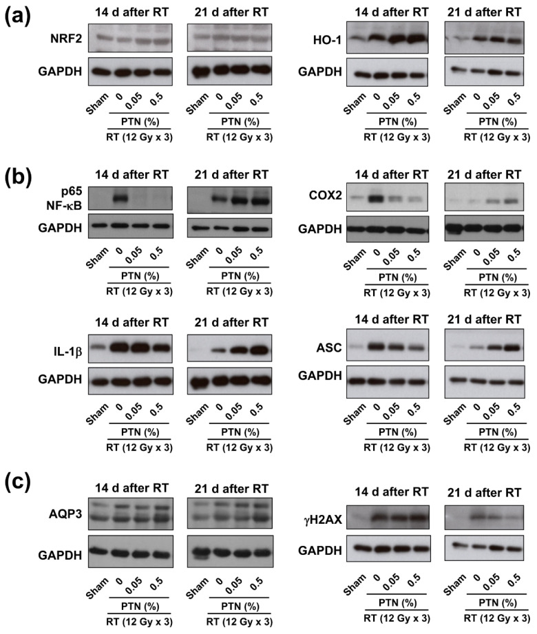
Radiation dermatitis (RD) is one of the most common side effects of radiotherapy; its symptoms progress from erythema to dry and moist desquamation, leading to the deterioration of the patients' quality of life. Active metabolites in brown seaweed, including phlorotannins (PTNs), show anti-inflammatory activities; however, their medical use is limited. Here, we investigated the effects of PTNs in a mouse model of RD in vivo. X-rays (36 Gy) were delivered in three fractions to the hind legs of BALB/c mice. Macroscopic RD scoring revealed that PTNs significantly mitigated RD compared with the vehicle control. Histopathological analyses of skin tissues revealed that PTNs decreased epidermal and dermal thickness compared with the vehicle control. Western blotting indicated that PTNs augmented nuclear factor erythroid 2-related factor 2 (NRF2)/heme oxygenase-1 (HO-1) pathway activation but attenuated radiation-induced NF-κB (nuclear factor kappa-light-chain-enhancer of activated B cells) and inflammasome activation, suggesting the mitigation of acute inflammation in irradiated mouse skin. PTNs also facilitated fast recovery, as indicated by increased aquaporin 3 expression and decreased γH2AX (histone family member X) expression. Our results indicate that topical PTN application may alleviate RD symptoms by suppressing oxidative stress and inflammatory signaling and by promoting the healing process. Therefore, PTNs may show great potential as cosmeceuticals for patients with cancer suffering from radiation-induced inflammatory side effects such as RD.

Keywords: inflammation; mouse model; phlorotannins; radiation dermatitis.


Conflict of interest statement

The authors declare no conflict of interest.

Figures

Figure 1
Figure 1
Radiation dermatitis scores in a BALB/c mouse model: (a) Definitions of the radiation dermatitis scores and (b) photographs of a mouse hind leg representing the radiation dermatitis scores.
Figure 2
Figure 2
Development of the mouse model of radiation dermatitis: (a) Time-course changes in the skin area irradiated with various radiation doses. Photographs were obtained at the indicated time points following irradiation. (b) Time-dependent scoring of radiation dermatitis: Data are shown as mean ± standard deviation (SD, n ≥ 5 per group). (c) Representative immunohistochemical staining images of the irradiated skin tissue sections: hematoxylin and eosin (upper panels) and Masson’s trichrome staining (bottom panels). Skin tissues were harvested at 21 days after irradiation. (d and e) Quantification data of the epidermis (d) and dermis (e) showed dose-dependent increases in their thickness. Data are shown as mean ± SD (n ≥ 80 per group). Difference was evaluated using a Kruskal–Wallis test, followed by Dunn’s multiple comparison test. ** p < 0.01; *** p < 0.001.
Figure 3
Figure 3
Effects of the topical application of phlorotannins (PTNs) on radiation dermatitis: (a) Schematic of the experimental procedures. Right hind legs were irradiated with X-rays at a dose of 12 Gy per day for three consecutive days (total, 36 Gy). PTNs were dissolved at the indicated concentrations in sesame oil and topically applied to the irradiated skin area starting on the irradiation day. An rhEGF (recombinant human epidermal growth factor) solution was used as the positive control. (b) Representative photographs of the irradiated skin treated with topical PTNs or rhEGF. (c) Time-course of changes in RD score: Data are shown as mean ± SD from three independent experiments. Difference was evaluated using one-way analysis of variance (ANOVA), followed by Bonferroni’s multiple comparison test. * p < 0.05; ** p < 0.01; *** p < 0.001.
Figure 4
Figure 4
Phlorotannins (PTNs) reduce the radiation-induced thickening of the epidermis and dermis. (a,b) Representative images of hematoxylin and eosin (H&E) and Masson’s trichrome staining of the irradiated skin tissues; The skin area was topically treated with 0%, 0.05%, and 0.5% PTN or EGF. The skin tissues were collected 14 (a) and 21 (b) days after irradiation. (c,d) Measurement of the epidermal and dermal thickness in the skin tissues collected 14 (c) and 21 (d) days after irradiation: Data are shown as mean ± SD. (n ≥ 40). Difference was evaluated using one-way ANOVA followed by Bonferroni’s multiple comparison test. ** p < 0.01; *** p < 0.001.
Figure 5
Figure 5
Phlorotannins (PTNs) mitigate radiation dermatitis by modulating the NRF2 and NF-κB signaling pathways: Skin tissue samples were collected 14 and 21 days after irradiation and subjected to western blotting. (a) Western blotting revealed that PTNs further enhanced the radiation-induced increase in NRF2/HO-1 (nuclear factor erythroid 2-related factor 2/heme oxygenase-1 pathway) expression on day 14. (b) PTNs attenuated the radiation-induced activation of the NF-κB pathway and inflammasome. (c) PTNs augmented radiation-induced aquaporin 3 (AQP3) expression but suppressed γH2AX expression in the irradiated tissues. Glyceraldehyde 3-phosphate dehydrogenase (GAPDH) was used as a loading control.

Similar articles

Cited by

References

    1. Hickok J.T., Morrow G.R., Roscoe J.A., Mustian K., Okunieff P. Occurrence, severity, and longitudinal course of twelve common symptoms in 1129 consecutive patients during radiotherapy for cancer. J. Pain Symptom Manag. 2005;30:433–442. doi: 10.1016/j.jpainsymman.2005.04.012. - DOI - PubMed
    1. Singh M., Alavi A., Wong R., Akita S. Radiodermatitis: A Review of Our Current Understanding. Am. J. Clin. Derm. 2016;17:277–292. doi: 10.1007/s40257-016-0186-4. - DOI - PubMed
    1. Lee J., Park W., Choi D.H., Huh S.J., Kim I.R., Kang D., Cho J. Patient-reported symptoms of radiation dermatitis during breast cancer radiotherapy: A pilot study. Qual. Life Res. 2017;26:1713–1719. doi: 10.1007/s11136-017-1526-4. - DOI - PubMed
    1. Pignol J.P., Olivotto I., Rakovitch E., Gardner S., Sixel K., Beckham W., Vu T.T., Truong P., Ackerman I., Paszat L. A multicenter randomized trial of breast intensity-modulated radiation therapy to reduce acute radiation dermatitis. J. Clin. Oncol. 2008;26:2085–2092. doi: 10.1200/JCO.2007.15.2488. - DOI - PubMed
    1. Wickline M.M. Prevention and treatment of acute radiation dermatitis: A literature review. Oncol. Nurs. Forum. 2004;31:237–247. doi: 10.1188/04.ONF.237-247. - DOI - PubMed

MeSH terms