. 2020 Jul 21;12(7):1989.
doi: 10.3390/cancers12071989.

Merkel Cell Polyomavirus T Antigens Induce Merkel Cell-Like Differentiation in GLI1-Expressing Epithelial Cells

Affiliations

Merkel Cell Polyomavirus T Antigens Induce Merkel Cell-Like Differentiation in GLI1-Expressing Epithelial Cells

Thibault Kervarrec et al. Cancers (Basel). .

Abstract

Merkel cell carcinoma (MCC) is an aggressive skin cancer frequently caused by the Merkel cell polyomavirus (MCPyV). It is still under discussion, in which cells viral integration and MCC development occurs. Recently, we demonstrated that a virus-positive MCC derived from a trichoblastoma, an epithelial neoplasia bearing Merkel cell (MC) differentiation potential. Accordingly, we hypothesized that MC progenitors may represent an origin of MCPyV-positive MCC. To sustain this hypothesis, phenotypic comparison of trichoblastomas and physiologic human MC progenitors was conducted revealing GLI family zinc finger 1 (GLI1), Keratin 17 (KRT 17), and SRY-box transcription factor 9 (SOX9) expressions in both subsets. Furthermore, GLI1 expression in keratinocytes induced transcription of the MC marker SOX2 supporting a role of GLI1 in human MC differentiation. To assess a possible contribution of the MCPyV T antigens (TA) to the development of an MC-like phenotype, human keratinocytes were transduced with TA. While this led only to induction of KRT8, an early MC marker, combined GLI1 and TA expression gave rise to a more advanced MC phenotype with SOX2, KRT8, and KRT20 expression. Finally, we demonstrated MCPyV-large T antigens' capacity to inhibit the degradation of the MC master regulator Atonal bHLH transcription factor 1 (ATOH1). In conclusion, our report suggests that MCPyV TA contribute to the acquisition of an MC-like phenotype in epithelial cells.

Keywords: ATOH1; GLI1; Merkel cell carcinoma; hair follicle; histogenesis; polyomavirus; sonic hedgehog.


Conflict of interest statement

The authors declare no conflict of interest.

Figures
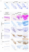
Figure 1
Figure 1
Merkel cells and possible Merkel cell progenitors in human skin. (A) Keratin 20 (KRT20), SRY-box transcription factor 2 (SOX2), KRT8, and KRT18 staining was used to identify Merkel cells (MCs) (bar = 100 µm) (only one hotspot investigated for illustration purpose). Merged analysis is available in Figure S1B. (B) Identification of potential MC progenitors in human skin: Three MC hotspots as well as interfollicular epidermis for comparison are depicted (bar = 100 µm) (15 hotspots investigated in total). Immunohistochemical staining revealed expression of KRT17 and SOX9 in the epidermal cells surrounding differentiated MCs suggesting that these cells are MC progenitors. Nuclear GLI family zinc finger 1 (GLI1) was detected only close to MC hotspots in hairy, but not in acral skin. Of note, neurofilament (NF)-expressing dermal nerves were observed in contact with the MCs.
Figure 2
Figure 2
Ectopic GLI1 expression in primary human epidermal keratinocytes induces several MC lineage markers: Normal human epidermal keratinocytes (NHEK) were infected with a lentiviral vector coding for GLI1 and puromycin resistance. Following antibiotic selection, cells were harvested after 14 days of cultivation. (A) Immunoblot analysis was performed to confirm GLI1 expression (insert), and isolated RNA was subjected to complementary DNA (cDNA) synthesis and real-time PCR. Relative messenger RNA (mRNA) expression levels of the indicated Merkel cell lineage markers are given as mean (+ standard error of the mean (SEM)) of four independent experiments (* p value < 0.05, paired t test) (mean CT value of the controls was used as reference). (B) Expression of GLI1, the MC progenitor (KRT17, SOX9) and the MC markers (SOX2, KRT8, and KRT20) was assessed by immunohistochemistry and relative protein expression quantification was performed on at least 1000 cells/condition using ImageJ software. Results are displayed as box and whiskers diagram with median, Q1, and Q3, as well as first and 99th percentile. These results were confirmed by two additional independent experiments (immunostaining and immunoblot) as shown in Figure S3. Uncropped membranes and Western blot signal quantifications are available in Figures S8 and S9, respectively.
Figure 3
Figure 3
T antigens induce expression of some early MC differentiation markers in primary human keratinocytes. A: NHEKs were infected with a lentiviral vector coding for small T (sT) and truncated Large T (LT) as well as a puromycin resistance. Following antibiotic selection, cells were analyzed after 14 days of cultivation. (A) Immunoblot analysis confirmed LT expression, and microscopic inspection revealed a less-flattened phenotype and cultures reaching much higher densities. Under microscopic examination such cells harbored reduced cytoplasmic size compared to the controls, as confirmed using imageJ software (bar = 100 µm) (* p value < 0.05, Mann–Whitney U test, n = 3 independent experiments). (B) Relative mRNA levels of the indicated Merkel cell differentiation markers (* p value < 0.05, paired t test, n = 4 independent experiments), (C) Immunoblot demonstrated T antigens (TA)-induced KRT8 protein expression and immunohistochemistry additionally revealed KRT8 expression is restricted to a subpopulation of small- to medium-sized round cells. Furthermore, occasionally “dot like” staining was observed (white arrows). (D) Immunohistochemical assessment of the indicated MC markers in TA-expressing NHEK, control NHEK and the MCC cell line WaGa (bar = 100 µm). KRT8 induction by T antigens was confirmed in two additional independent experiments, which are depicted in Figure S4. For relative quantification of protein expression levels, at least 1000 cells/condition were evaluated using ImageJ software. Results are displayed as box and whiskers diagram with median, Q1, and Q3 as well as first and 99th percentile. Uncropped membranes and Western blot signal quantifications are available in Figures S8 and S9, respectively.
Figure 4
Figure 4
Induction of late MC markers by combined expression of GLI1 and Merkel cell Polyomavirus (MCPyV) T antigens (TA) in primary keratinocytes. NHEKs were infected with a bicistronic lentiviral vector coding for GLI1 as well as sT and truncated LT. Under control of a second promoter, a pure resistance was expressed. Following antibiotic selection, cells were analyzed after 14 days of cultivation. (A) GLI1/TA combined ectopic expression was associated with formation of floating clusters of living cells in normal human epidermal keratinocytes (NHEK), while these findings were not observed in controls or when GLI1 and TA were transduced independently (PC: Phase contrast) (Figure S4) (n = 3 independent experiments). White arrows indicate the floating cells. (B,C) Immunohistochemical assessment of Merkel cell markers (SOX2, KRT8, and KRT20) expression levels in GLI1/T antigen-expressing NHEKs and controls. Immunohistochemistry was performed on the respective cells spotted on slides (2 × 10⁵ cells/condition). B. Count of cells expressing the Merkel cell markers in GLI1/T antigens (TA)-expressing NHEK and controls (results are mean ±SEM of three independent experiments). Counting of positive cells was preferred to relative protein level quantification due to the low number of GLI1/TA-expressing cells. C. Representative photos of LT, GLI1, SOX2, KRT8, KRT18, and KRT20 expression in NHEK (controls), GLI1/TA-expressing NHEK, and the WaGa MCC cell line. White arrows indicate cells expressing the respective proteins. The results for two additional independent experiments are shown in Figure S5.
Figure 5
Figure 5
MCPyV T antigens increase the half-life of ATOH1 (A) Hemagglutinin (HA)-tagged ATOH1- and/or TA-encoding plasmids were transfected either individually or combined into U2OS cells. After two days, real-time PCR and immunoblot analyses were performed. While ATOH1 mRNA was not affected (mean ± SEM of three independent experiments), ATOH1 protein accumulation in the presence of TA was observed. (B) Co-transfection of a constant amount (0.3 µg) of HA-tagged ATOH1 and increasing amounts of TA in U2OS cells followed by immunoblot analysis. ATOH1-HA signals relative to actin were quantified using ImageJ. Mean ± SEM of three independent experiments was displayed. (C) Evaluation of ATOH1 half-life in absence or presence of T antigens. Twenty-four hours after transfection, HEK293 cells were exposed to the translation inhibitor cycloheximide (CHX) for variable durations (0–6 h). ATOH1-HA expression was then evaluated by immunoblot analysis and quantified using the Image J Software (mean ± SEM of three independent experiments are depicted). (D) A mutant of ATOH1-HA, in which the three serines at positions 331, 337, and 342 were all exchanged to alanines (ATOH1-HA-3A), was generated, and the impact of co-transfected TA on ATOH1-HA wild type and ATOH1-HA-3A expression was analyzed in CHX chase experiments (see C). Quantified signals relative to actin are given in the graphs below. (E) Co-transfection of ATOH1-HA-3A with increasing amounts of TA did not affect ATOH1 protein expression level (this was confirmed in a second independent experiment). Uncropped membranes and Western blot signal quantifications are available in Figures S8 and S9, respectively.

References

    1. Lemos B.D., Storer B.E., Iyer J.G., Phillips J.L., Bichakjian C.K., Fang L.C., Johnson T.M., Liegeois-Kwon N.J., Otley C.C., Paulson K.G., et al. Pathologic nodal evaluation improves prognostic accuracy in Merkel cell carcinoma: Analysis of 5823 cases as the basis of the first consensus staging system. J. Am. Acad. Dermatol. 2010;63:751–761. doi: 10.1016/j.jaad.2010.02.056. - DOI - PMC - PubMed
    1. Feng H., Shuda M., Chang Y., Moore P.S. Clonal integration of a polyomavirus in human Merkel cell carcinoma. Science. 2008;319:1096–1100. doi: 10.1126/science.1152586. - DOI - PMC - PubMed
    1. Verhaegen M.E., Mangelberger D., Harms P.W., Eberl M., Wilbert D.M., Meireles J., Bichakjian C.K., Saunders T.L., Wong S.Y., Dlugosz A.A. Merkel cell polyomavirus small T antigen initiates Merkel cell carcinoma-like tumor development in mice. Cancer Res. 2017;77:3151–3157. doi: 10.1158/0008-5472.CAN-17-0035. - DOI - PMC - PubMed
    1. Tilling T., Moll I. Which are the cells of origin in merkel cell carcinoma? J. Skin Cancer. 2012;2012:680410. doi: 10.1155/2012/680410. - DOI - PMC - PubMed
    1. Becker J.C., Zur Hausen A. Cells of origin in skin cancer. J. Investig. Dermatol. 2014;134:2491–2493. doi: 10.1038/jid.2014.233. - DOI - PubMed