. 2020 Jul 21;6(3):112.
doi: 10.3390/jof6030112.

Copper-Chitosan Nanocomposite Hydrogels Against Aflatoxigenic Aspergillus flavus from Dairy Cattle Feed

Affiliations

Copper-Chitosan Nanocomposite Hydrogels Against Aflatoxigenic Aspergillus flavus from Dairy Cattle Feed

Kamel A Abd-Elsalam et al. J Fungi (Basel). .

Abstract

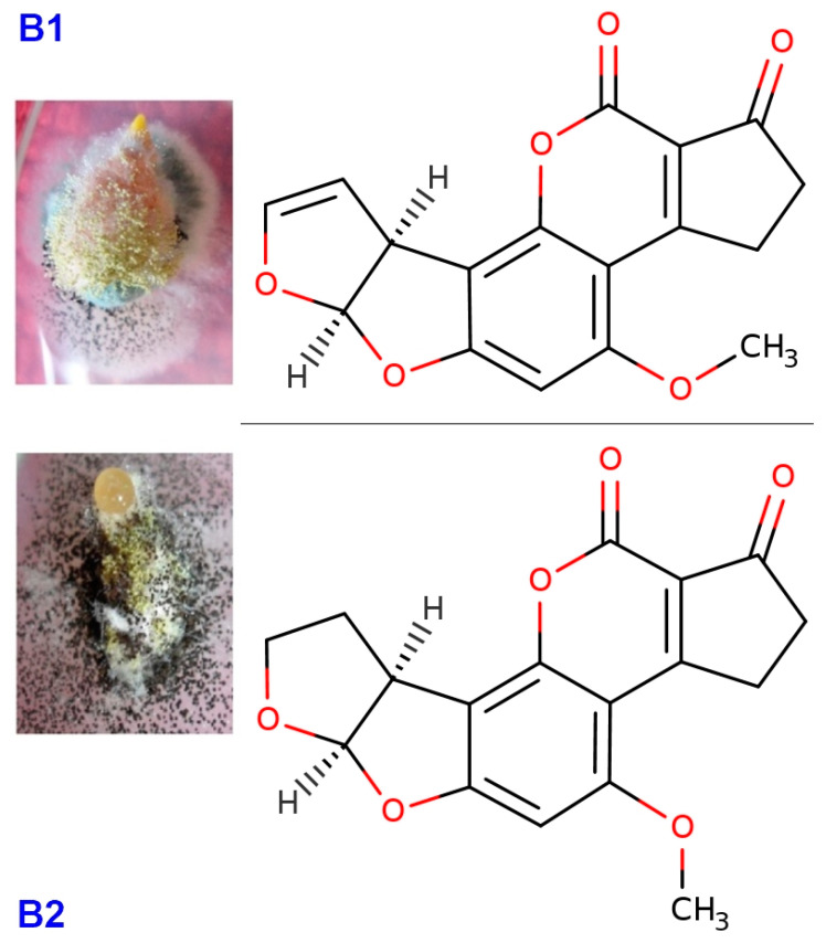
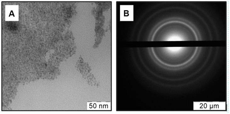
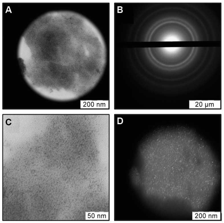
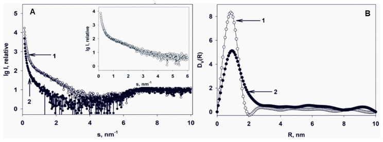
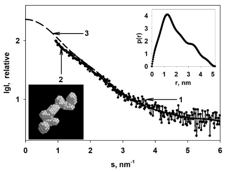
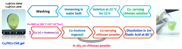
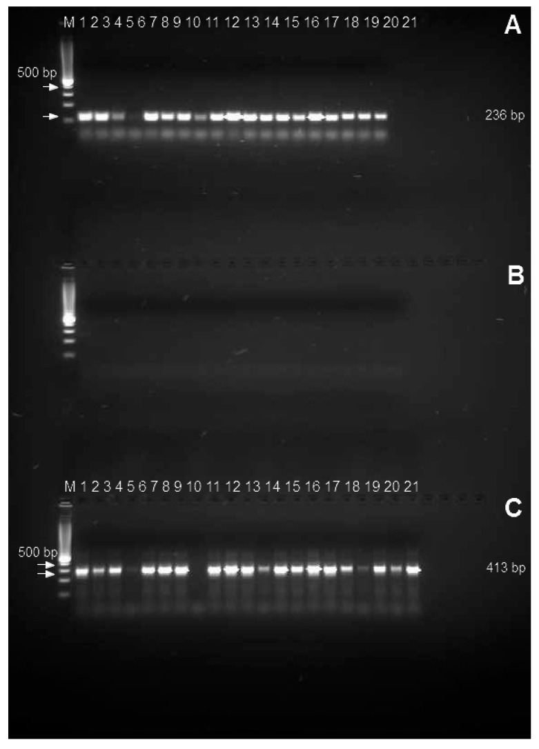
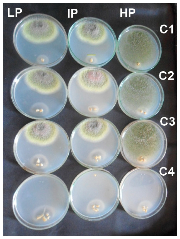
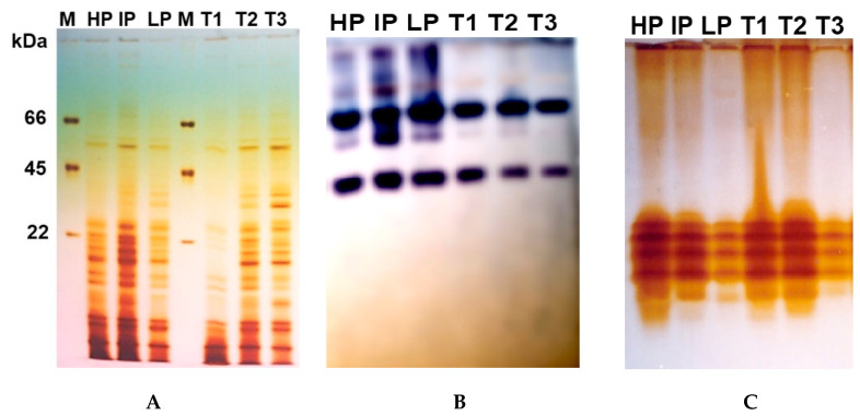
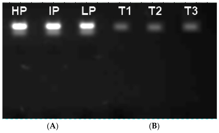
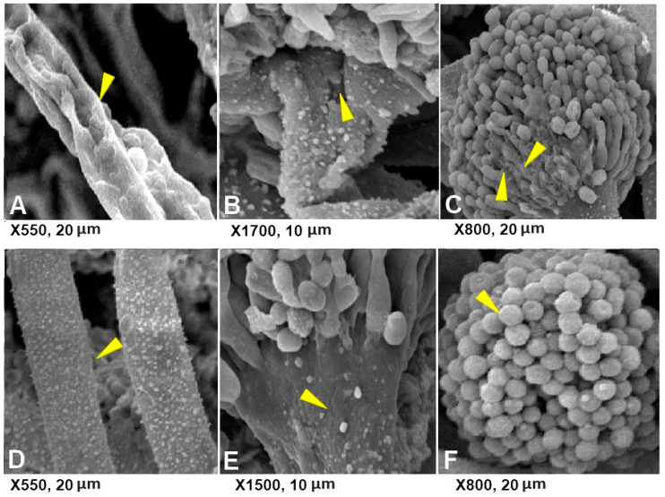
The integration of copper nanoparticles as antifungal agents in polymeric matrices to produce copper polymer nanocomposites has shown excellent results in preventing the growth of a wide variety of toxigenic fungi. Copper-chitosan nanocomposite-based chitosan hydrogels (Cu-Chit/NCs hydrogel) were prepared using a metal vapor synthesis (MVS) and the resulting samples were described by transmission electron microscopy (TEM), X-ray fluorescence analysis (XRF), and small-angle X-ray scattering (SAXS). Aflatoxin-producing medium and VICAM aflatoxins tests were applied to evaluate their ability to produce aflatoxins through various strains of Aspergillus flavus associated with peanut meal and cotton seeds. Aflatoxin production capacity in four fungal media outlets revealed that 13 tested isolates were capable of producing both aflatoxin B1 and B2. Only 2 A. flavus isolates (Af11 and Af 20) fluoresced under UV light in the A. flavus and parasiticus Agar (AFPA) medium. PCR was completed using two specific primers targeting aflP and aflA genes involved in the synthetic track of aflatoxin. Nevertheless, the existence of aflP and aflA genes indicated some correlation with the development of aflatoxin. A unique DNA fragment of the expected 236 bp and 412 bp bands for aflP and aflA genes in A. flavus isolates, although non-PCR fragments have been observed in many other Aspergillus species. This study shows the antifungal activity of Cu-Chit/NCs hydrogels against aflatoxigenic strains of A. flavus. Our results reveal that the antifungal activity of nanocomposites in vitro can be effective depending on the type of fungal strain and nanocomposite concentration. SDS-PAGE and native proteins explain the apparent response of cellular proteins in the presence of Cu-Chit/NCs hydrogels. A. flavus treated with a high concentration of Cu-Chit/NCs hydrogels that can decrease or produce certain types of proteins. Cu-Chit/NCs hydrogel decreases the effect of G6DP isozyme while not affecting the activity of peroxidase isozymes in tested isolates. Additionally, microscopic measurements of scanning electron microscopy (SEM) showed damage to the fungal cell membranes. Cu-Chit/NCS hydrogel is an innovative nano-biopesticide produced by MVS is employed in food and feed to induce plant defense against toxigenic fungi.

Keywords: Aspergillus section Flavi; aflatoxins; chitosan; feeds; nanocomposites.


Conflict of interest statement

The authors declare no conflict of interest. The funders had a main role in the design of the study; in the collection, analyses, or interpretation of data; in the writing of the manuscript, or in the decision to publish the results.

Figures

Figure 1
Figure 1
Structural formula of two type of aflatoxins produced by A. flavus isolated from peanut meal and cotton seed. Aflatoxin (B1) C17H12O6 (6aR,9aS)-4-Methoxy-2,3,6a,9a-tetrahydrocyclopenta[c]furo[3′,2′:4,5]furo[2,3-h]chromen-1,11-dion. Aflatoxin (B2), C17H14O6 (6aS,9aR)-4-Methoxy-2,3,6a,8,9,9a hexahydrocyclopenta[c]furo[3′,2′:4,5]furo[2,3-h]chromen-1,11-dion. Formula available online from: http://www.chemspider.com.
Figure 2
Figure 2
TEM image in bright field (A) and selected area diffraction pattern (SAED) (B) from a region for Cu-acetone organosol.
Figure 3
Figure 3
TEM images in bright (A,C) and dark fields (D) of chitosan with a high molecular weight (ChitHMW) doped with copper nanoparticles (Cu NPs) as well as SAED (B) of highlighted field.
Figure 4
Figure 4
(A) Experimental (SAXS) curves: 1—Cu-carrying chitosan; 2—pristine non-modified chitosan (chitosan low molecular weight (ChitLMW)). Insert—difference SAXS curve for the embedded Cu nanoparticles. (B) Volume size distribution functions DV(R): 1—Cu nanoparticles; 2—pristine non-modified chitosan (ChitLMW).
Figure 5
Figure 5
Reconstruction of the shape of the Cu nanoparticles in the Cu-carrying chitosan: 1—difference SAXS curve; 2—a model scattering curve calculated from the restored shape of the Cu nanoparticles; 3—extrapolated to zero angles smoothed scattering curve after the introduction of collimation corrections. Inserts: top right—distance distribution function p(r); bottom left—restored shape of the Cu nanoparticles.
Figure 6
Figure 6
Scheme of preparation of chitosan hydrogels from Cu-carrying chitosan powders.
Figure 7
Figure 7
PCR amplicons obtained using primer pairs developed for the aflP (omtA) and aflA genes in 21 Aspergillus flavus (Lane1–21) tested with aflP primers (A), and an Aspergillus related isolate such as two isolates for both of Aspergillus clavatus, Aspergillus ochraceous, Aspergillus niveus, Aspergillus terreus, Aspergillus fumigatus, Aspergillus versicolor, Penicillium paneum, Penicillium expansum, Penicillium citrinum, Penicillium verrucosum and one Alternaria alternate isolate (Lane 1–21) tested with aflP primers (B). A total of 21 Aspergillus flavus (Lane1–21) tested with aflA primers (C).
Figure 8
Figure 8
Antifungal activity for different concentration of Cu-Chit/NCs gel (C1 = 60, C2 = 120, C3 = 180, and C4 = 240 ppm) against A. flavus isolated from feeds by the plate assay. All Petri dish treatments were incubated at 28 °C for one week.
Figure 9
Figure 9
(A) Protein expression profile of SDS-PAGE extracted from A. flavus mycelium treated with a high Cu-Chit/NCs gel concentration. Lane M shows a standard protein molecular weight marker. Protein marker including three molecular bands ranging from 66, 45, and 22 kDa was used. Isoenzymes electrophoresis of G6PD (B) and peroxidase (C) isozymes extracted from A. flavus mycelium treated with a high Cu-Chit/NCs gel concentration.
Figure 10
Figure 10
Agarose gel electrophoretic pattern of the fungal genomic DNA treated with 180 ppm of Cu-Chit/NCs gel, (A) Lanes 1–3: DNA for untreated A. flavus isolates, Lane 1: A. flavus (high producer (HP) isolate), Lane 2: A. flavus (intermediate producer (IP) isolate), Lane 3: A. flavus (low producer (LP) isolate). (B) Lanes T1, T2, and T3, three A. flavus isolates DNA treated with Cu-Chit/NCs gel, showed total damage to fragmented DNA bands.
Figure 11
Figure 11
Scanning electron micrographs showing shriveled conidiophores (AC) and the healthy mycelium or conidiophores (DF) of A. flavus on PDA treated with Cu-Chit/NCs hydrogel. The yellow arrows mean hyphae or conidiophore.

References

    1. Almoammar H., Bahkali A.H., Abd-Elsalam K.A. A Polyphasic method for the identification of aflatoxigenic’aspergillus’ species isolated from camel feeds. Aust. J. Crop. Sci. 2013;7:1707.
    1. Omeiza G.K., Kabir J., Kwaga J.K.P., Kwanashie C.N., Mwanza M., Ngoma L. A Risk assessment study of the occurrence and distribution of aflatoxigenic Aspergillus flavus and Aflatoxin B1 in dairy cattle feeds in a Central Northern State, Nigeria. Toxicol. Rep. 2018;5:846–856. doi: 10.1016/j.toxrep.2018.08.011. - DOI - PMC - PubMed
    1. Li Z., Lin S., An S., Liu L., Hu Y., Wan L. Preparation, characterization and Anti-Aflatoxigenic activity of chitosan packaging films incorporated with turmeric essential oil. Int. J. Biol. Macromol. 2019;131:420–434. doi: 10.1016/j.ijbiomac.2019.02.169. - DOI - PubMed
    1. Mateo E.M., Gómez J.V., Domínguez I., Gimeno-Adelantado J.V., Mateo-Castro R., Gavara R., Jiménez M. Impact of bioactive packaging systems based on EVOH films and essential oils in the control of aflatoxigenic fungi and aflatoxin production In Maize. Int. J. Food Microbiol. 2017;254:36–46. doi: 10.1016/j.ijfoodmicro.2017.05.007. - DOI - PubMed
    1. Kumar A., Singh P.P., Prakash B. Unravelling the antifungal and Anti-Aflatoxin B1 Mechanism of Chitosan Nanocomposite Incorporated with Foeniculum vulgare essential oil. Carbohydr. Polym. 2020;236:116050. doi: 10.1016/j.carbpol.2020.116050. - DOI - PubMed

LinkOut - more resources