Review
. 2020 Jul 21;9(7):2319.
doi: 10.3390/jcm9072319.

Surgery and Perioperative Management in Small Intestinal Neuroendocrine Tumors

Affiliations
Review

Surgery and Perioperative Management in Small Intestinal Neuroendocrine Tumors

Sophie Deguelte et al. J Clin Med. .

Abstract

Small-intestinal neuroendocrine tumors (SI-NETs) are the most prevalent small bowel neoplasms with an increasing frequency. In the multimodal management of SI-NETs, surgery plays a key role, either in curative intent, even if R0 resection is feasible in only 20% of patients due to advanced stage at diagnosis, or palliative intent. Surgeons must be informed about the specific surgical management of SI-NETs according to their hormonal secretion, their usual dissemination at the time of diagnosis and the need for bowel-preserving surgery to avoid short bowel syndrome. The aim of this paper is to review the surgical indications and techniques, and perioperative and postoperative management of SI-NETs.

Keywords: carcinoid crisis; carcinoid syndrome; small bowel neuroendocrine tumor; surgery.


Conflict of interest statement

Board AAA, IPSEN, Novartis, Keocyt GC; board Novartis SD.

Figures

Figure 1
Figure 1
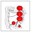
Classical presentation of small-intestinal neuroendocrine tumors (SI-NETs). (a) Small primary tumors (<20 mm), distal in the ileum, and multiple in 30% to 50% of cases; (b) Mesenteric lymph node metastases present in more than 80% of cases at diagnosis, typically larger than primary tumors; (c) Liver metastases present in approximately 50% of cases at diagnosis usually multiple and bilobar.
Figure 2
Figure 2
Classification based on the relationship between mesenteric lymph node metastases (MLNM) and the superior mesenteric vessels. Lymph node stage 0: no visible mesenteric MLNM suspicious of malignancy; Lymph node stage I: proximity to the small intestine, without invasion of the superior mesenteric artery lymph node (LN); Lymph node stage II: involvement of the distal branches of the superior mesenteric artery (SMA), next to their origin; Lymph node stage III: involvement of the trunk of the SMA, without involving the first jejunal arteries; III ‘up’: <3–4 free jejunal branches; III ‘down’: >3–4 free jejunal branches; Lymph node stage IV: involvement of the trunk of the SMA with involvement of the first jejunal arteries.
Figure 3
Figure 3
Surgical pattern: (a) no more ‘pizza pie’ approach: large intestinal resection with inappropriate lymphadenectomy. (b) Appropriate lymphadenectomy should remove at least 8 (better 12) lymph nodes with small intestinal resection. The ileocecal valve and right colon might require resection mainly for LN-stage III patients.
Figure 4
Figure 4
Operative view during the lymphadenectomy. (a) Clamping test after the dissection of the mesenteric superior artery and before the resection of the small bowel to visualize the remnant vascularized bowel. The blue arrow shows the mesenteric superior artery, and the green arrow shows the firsts jejunal arteries. (b) Small bowel vascularized by the remnant jejunal arteries.
Figure 5
Figure 5
Mesenteric ischemia due to bulky MLNM with encasement of the mesenteric vessels. (a) Computed tomography (CT) scan: mesenteric superior venous stenosis (blue arrow) and MLNM (green arrow). (b) Debulking surgery for mesenteric ischemia due to mesenteric vessels encasement by MLNM.
Figure 6
Figure 6
Operative view of a hybrid procedure combining laparoscopy and manual palpation of the entire small bowel. (a) Laparoscopic preparation; (b) Exteriorization of all the entire small bowel, the right colon, and the mesentery; (c) Postoperative scarce.
Figure 7
Figure 7
Proposed algorithm diagram for surgical indications of resection for SI-NET local disease. * Resection of all the primary tumors (after manual palpation of the entire small bowel) + systematic mesenteric lymphadenectomy (with at least 8 or 12 removed LN).

References

    1. Dasari A., Shen C., Halperin D., Zhao B., Zhou S., Xu Y., Shih T., Yao J.C. Trends in the Incidence, Prevalence, and Survival Outcomes in Patients with Neuroendocrine Tumors in the United States. JAMA Oncol. 2017;3:1335–1342. doi: 10.1001/jamaoncol.2017.0589. - DOI - PMC - PubMed
    1. Pape U.-F., Berndt U., Muller-Nordhorn J., Bohmig M., Roll S., Koch M., Willich S.N., Wiedenmann B. Prognostic factors of long-term outcome in gastroenteropancreatic neuroendocrine tumours. Endocr. Relat. Cancer. 2008;15:1083–1097. doi: 10.1677/ERC-08-0017. - DOI - PubMed
    1. Norlén O., Stålberg P., Öberg K., Eriksson J., Hedberg J., Hessman O., Janson E.T., Hellman P., Åkerström G. Long-Term Results of Surgery for Small Intestinal Neuroendocrine Tumors at a Tertiary Referral Center. World J. Surg. 2012;36:1419–1431. doi: 10.1007/s00268-011-1296-z. - DOI - PubMed
    1. Strosberg J.R., Weber J.M., Feldman M., Coppola D., Meredith K., Kvols L.K. Prognostic validity of the American Joint Committee on Cancer staging classification for midgut neuroendocrine tumors. J. Clin. Oncol. 2013;31:420–425. doi: 10.1200/JCO.2012.44.5924. - DOI - PubMed
    1. Le Roux C., Lombard-Bohas C., Delmas C., Dominguez-Tinajero S., Ruszniewski P., Samalin E., Raoul J.-L., Renard P., Baudin E., Robaskiewicz M., et al. Relapse factors for ileal neuroendocrine tumours after curative surgery: A retrospective French multicentre study. Dig. Liver Dis. 2011;43:828–833. doi: 10.1016/j.dld.2011.04.021. - DOI - PubMed

LinkOut - more resources