. 2020 Jul 18;9(7):1726.
doi: 10.3390/cells9071726.

miR-22-3p Negatively Affects Tumor Progression in T-Cell Acute Lymphoblastic Leukemia

Affiliations

miR-22-3p Negatively Affects Tumor Progression in T-Cell Acute Lymphoblastic Leukemia

Valentina Saccomani et al. Cells. .

Abstract

T-cell acute lymphoblastic leukemia (T-ALL) is a rare, aggressive disease arising from T-cell precursors. NOTCH1 plays an important role both in T-cell development and leukemia progression, and more than 60% of human T-ALLs harbor mutations in components of the NOTCH1 signaling pathway, leading to deregulated cell growth and contributing to cell transformation. Besides multiple NOTCH1 target genes, microRNAs have also been shown to regulate T-ALL initiation and progression. Using an established mouse model of T-ALL induced by NOTCH1 activation, we identified several microRNAs downstream of NOTCH1 activation. In particular, we found that NOTCH1 inhibition can induce miR-22-3p in NOTCH1-dependent tumors and that this regulation is also conserved in human samples. Importantly, miR-22-3p overexpression in T-ALL cells can inhibit colony formation in vitro and leukemia progression in vivo. In addition, miR-22-3p was found to be downregulated in T-ALL specimens, both T-ALL cell lines and primary samples, relative to immature T-cells. Our results suggest that miR-22-3p is a functionally relevant microRNA in T-ALL whose modulation can be exploited for therapeutic purposes to inhibit T-ALL progression.

Keywords: NOTCH1; T-ALL; miR-22-3p.


Conflict of interest statement

The authors declare no conflicts of interest.

Figures

Figure 1
Figure 1
Heat map of significantly regulated miRNAs following NOTCH1 inhibition in vivo. MiRNA expression profiling was performed in three biological replicates of an HD-ΔPEST-NOTCH1 T-cell tumor treated in vivo with vehicle (DMSO) or with 5 mg/kg of dibenzazepine (DBZ). The heat map shows the significantly differentially expressed miRNAs (|FC| > 1.3; FDR < 0.05). A hierarchical clustering was performed using the Euclidean distance and the complete linkage method on both samples (columns) and miRNAs (rows). The unsupervised analysis clearly separated two clusters representing downregulated miRNAs (top) and upregulated miRNAs (down) in DBZ-treated vs. control.
Figure 2
Figure 2
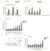
Anti-NOTCH1 treatment in murine and human T-ALL cells results in miR-22-3p upregulation. (A,B) qPCR analysis of miR-22-3p expression upon DBZ treatment in murine models of NOTCH1-induced T-ALLs: (A) leukemic cells obtained from three mice transplanted with the HD-∆PEST-NOTCH1-tumor-1 (T1), treated in vivo with DMSO or DBZ, were analyzed; (B) five independent ∆E-NOTCH1-tumors (T1-5), treated in vivo with DMSO or DBZ, were analyzed. (C) qPCR analysis of miR-22-3p expression upon DBZ treatment of T-ALL cell lines (treated in vitro with DBZ for 72 h). (D) Western blot analysis of intracellular cleaved NOTCH1 (ICN1) in PDX samples treated in vivo with vehicle only (DMSO) or with 5 mg/kg DBZ, both pre- and post-depletion. α-Tubulin was used as loading control. (E) The depleted PDX samples were analyzed by qPCR for miR-22-3p expression. Data are represented as mean ± SD. Assays were performed in triplicate (* p < 0.05, ** p < 0.01, and *** p < 0.001).
Figure 3
Figure 3
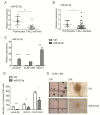
miR-22-3p is downregulated in T-ALL cells and negatively affects colony formation. (A,B) qPCR analysis of miR-22-3p expression in T-ALL cell lines (n = 17) and primary T-ALL samples (n = 30) compared with normal thymocytes (n = 7). (C) qPCR analysis of miR-22-3p overexpression in Jurkat-E6, CCRF-CEM, and MOLT4 cell lines. (D,E) T-cell lines showing the effects of miR-22-3p overexpression in colony-forming unit (CFU) assays using methylcellulose-based medium (* p < 0.05, ** p < 0.01, and *** p < 0.001). Representative images of CCRF-CEM colonies using an inverted microscope at different magnifications (10× and 20×). Data are represented as mean ± SD. Assays were performed in triplicate. (* p < 0.05, ** p < 0.01, and *** p < 0.001.).
Figure 4
Figure 4
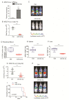
miR-22-3p overexpression in T-ALL cells delays tumor growth in vivo. (A) MOLT4 cells expressing luciferase were engineered to stably express miR-22-3p or empty vector (ctrl). (B,C) miR-22-3p or empty vector control MOLT4 cells were injected in vivo in NOD.Cg-Prkdcscid Il2rgtm1Wjl/SzJ (NSG) mice (n = 6). Bioluminescence quantification and representative images of NSG mice at 14 days from injection (*** p < 0.001). (DF) Flow cytometry analysis of human CD45 expression in the peripheral blood, in the spleen, and in the liver at day 21 after injection (n = 6 Ctrl vs. n = 6 miR-22-3p overexpressing group; ** p < 0.01 and *** p < 0.001). (GI) Bioluminescence quantification and representative images of NSG mice at day 22 after injection (n = 10 Ctrl vs. n = 10 miR-22-3p-overexpressing group). Overall survival is shown in (I) (*** p < 0.001).
Figure 5
Figure 5
miR-22-3p overexpression in T-ALL cells leads to the downregulation of multiple NOTCH1 targets. (A) qPCR analysis of human MYC, HES1, DTX1, PTCRA IGF1R, and NOTCH3 in MOLT4, Jurkat-E6, and CUTLL1 cells engineered to stably express miR-22-3p or empty vector (Ctrl) (* p < 0.05, ** p < 0.01, and *** p < 0.001). (B) Western blot analysis of HES1 and IGF1R in MOLT4 cells overexpressing miR-22-3p. α-Tubulin was used as loading control. Data are represented as mean ± SD.

References

    1. Pui C.H., Carroll W.L., Meshinchi S., Arceci R.J. Biology, risk stratification, and therapy of pediatric acute leukemias: An update. J. Clin. Oncol. Off. J. Am. Soc. Clin. Oncol. 2011;29:551–565. doi: 10.1200/JCO.2010.30.7405. - DOI - PMC - PubMed
    1. Weng A.P., Ferrando A.A., Lee W., Morris J.P.T., Silverman L.B., Sanchez-Irizarry C., Blacklow S.C., Look A.T., Aster J.C. Activating mutations of NOTCH1 in human T cell acute lymphoblastic leukemia. Science. 2004;306:269–271. doi: 10.1126/science.1102160. - DOI - PubMed
    1. Pear W.S., Aster J.C., Scott M.L., Hasserjian R.P., Soffer B., Sklar J., Baltimore D. Exclusive development of T cell neoplasms in mice transplanted with bone marrow expressing activated Notch alleles. J. Exp. Med. 1996;183:2283–2291. doi: 10.1084/jem.183.5.2283. - DOI - PMC - PubMed
    1. Deftos M.L., Huang E., Ojala E.W., Forbush K.A., Bevan M.J. Notch1 signaling promotes the maturation of CD4 and CD8 SP thymocytes. Immunity. 2000;13:73–84. doi: 10.1016/S1074-7613(00)00009-1. - DOI - PMC - PubMed
    1. Gonzalez-Garcia S., Garcia-Peydro M., Martin-Gayo E., Ballestar E., Esteller M., Bornstein R., de la Pompa J.L., Ferrando A.A., Toribio M.L. CSL-MAML-dependent Notch1 signaling controls T lineage-specific IL-7R{alpha} gene expression in early human thymopoiesis and leukemia. J. Exp. Med. 2009;206:779–791. doi: 10.1084/jem.20081922. - DOI - PMC - PubMed

Publication types

MeSH terms

LinkOut - more resources