Review
. 2020 Jul 16;11(7):809.
doi: 10.3390/genes11070809.

Epigenetics as an Evolutionary Tool for Centromere Flexibility

Affiliations
Review

Epigenetics as an Evolutionary Tool for Centromere Flexibility

Laura Leo et al. Genes (Basel). .

Abstract

Centromeres are the complex structures responsible for the proper segregation of chromosomes during cell division. Structural or functional alterations of the centromere cause aneuploidies and other chromosomal aberrations that can induce cell death with consequences on health and survival of the organism as a whole. Because of their essential function in the cell, centromeres have evolved high flexibility and mechanisms of tolerance to preserve their function following stress, whether it is originating from within or outside the cell. Here, we review the main epigenetic mechanisms of centromeres' adaptability to preserve their functional stability, with particular reference to neocentromeres and holocentromeres. The centromere position can shift in response to altered chromosome structures, but how and why neocentromeres appear in a given chromosome region are still open questions. Models of neocentromere formation developed during the last few years will be hereby discussed. Moreover, we will discuss the evolutionary significance of diffuse centromeres (holocentromeres) in organisms such as nematodes. Despite the differences in DNA sequences, protein composition and centromere size, all of these diverse centromere structures promote efficient chromosome segregation, balancing genome stability and adaptability, and ensuring faithful genome inheritance at each cellular generation.

Keywords: CENP-A; centromere; centromere evolution; holocentromere; neocentromere; repetitive sequences.


Conflict of interest statement

The authors declare no conflict of interest.

Figures

Figure 1
Figure 1
Schematic representation of the functional role of Ash1, CBP, and Trx proteins in the centromeric region. These proteins work by modifying the epigenetic state of the centromeric chromatin. In particular, on H3 histones, Ash1 dimethylates lysine 4 (H3K4me2) and CBP acetylates lysine 27 (H3K27ac) within the centromeric region. Trx works by inducing chromatin opening which, in turn, favours both CENP-A/Cid deposition and activation of transcription.
Figure 2
Figure 2
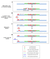
Schematic representation of the principal models of neocentromere formation: (A) Spreading model and activation model: centromeric markers spread from centromere into adjacent areas inducing neocentromere formation near endogenous centromeres following a DNA double-strand break (DSB) (B) Latent centromere model and inhibition lateral model: ectopic sites along a chromosome have an intrinsic ability to perform centromeric activity, but they are repressed by a dominant centromere. When the endogenous centromere is inactivated, one of these sites becomes active and functionally competent. (C) DNA breaking model: a neocentromere emerges at a breaking site where centromeric protein A (CENP-A) is rapidly recruited. (D) CENP-A island model: CENP-A is deposited on islands of nucleosomes scattered along the chromosomes. A random insertion of a transposable element (TE) in a CENP-A-ectopic site represents the first step towards the formation of a new, fully functional centromere.
Figure 3
Figure 3
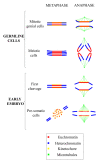
Diagrammatic representation of the variations in the centromere position and chromatin and kinetic activity in different cell types. Note, kinetic activity is retained exclusively by euchromatin in embryonic presomatic cells, in gonial cells by the entire chromosome and in meiotic cells only by telomeric heterochromatin.

References

    1. Karpen G.H., Allshire R.C. The case for epigenetic effects on centromere identity and function. Trends Genet. 1997;13:489–496. doi: 10.1016/S0168-9525(97)01298-5. - DOI - PubMed
    1. Earnshaw W.C., Rothfield N. Identification of a family of human centromere proteins using autoimmune sera from patients with scleroderma. Chromosoma. 1985;91:313–321. doi: 10.1007/BF00328227. - DOI - PubMed
    1. Sullivan K.F., Hechenberger M., Masri K. Human CENP-A contains a histone H3 related histone fold domain that is required for targeting to the centromere. J. Cell Biol. 1994;127:581–592. doi: 10.1083/jcb.127.3.581. - DOI - PMC - PubMed
    1. Buchwitz B.J., Ahmad K., Moore L.L., Roth M.B., Henikoff S. A histone-H3-like protein in C. elegans. Nature. 1999;401:547–548. doi: 10.1038/44062. - DOI - PubMed
    1. Drinnenberg I.A., DeYoung D., Henikoff S., Malik H.S. Recurrent loss of CenH3 is associated with independent transitions to holocentricity in insects. ELife. 2014;3:e03676. doi: 10.7554/eLife.03676. - DOI - PMC - PubMed

Publication types

LinkOut - more resources