. 2020 Jul 17;25(14):3267.
doi: 10.3390/molecules25143267.

Effect of Quercetin on Dexamethasone-Induced C2C12 Skeletal Muscle Cell Injury

Affiliations

Effect of Quercetin on Dexamethasone-Induced C2C12 Skeletal Muscle Cell Injury

Chun Chen et al. Molecules. .

Abstract

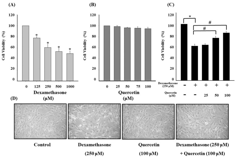
Glucocorticoids are widely used anti-inflammatory drugs in clinical settings. However, they can induce skeletal muscle atrophy by reducing fiber cross-sectional area and myofibrillar protein content. Studies have proven that antioxidants can improve glucocorticoid-induced skeletal muscle atrophy. Quercetin is a potent antioxidant flavonoid widely distributed in fruits and vegetables and has shown protective effects against dexamethasone-induced skeletal muscle atrophy. In this study, we demonstrated that dexamethasone significantly inhibited cell growth and induced cell apoptosis by stimulating hydroxyl free radical production in C2C12 skeletal muscle cells. Our results evidenced that quercetin increased C2C12 skeletal cell viability and exerted antiapoptotic effects on dexamethasone-treated C2C12 cells by regulating mitochondrial membrane potential (ΔΨm) and reducing oxidative species. Quercetin can protect against dexamethasone-induced muscle atrophy by regulating the Bax/Bcl-2 ratio at the protein level and abnormal ΔΨm, which leads to the suppression of apoptosis.

Keywords: C2C12 skeletal muscle cells; antioxidant; apoptosis; dexamethasone; mitochondrial membrane potential (ΔΨm); quercetin.


Conflict of interest statement

The authors declare no conflict of interest.

Figures

Figure 1
Figure 1
(A) Cell viability of C2C12 myotube cells in the presence of 0, 125, 250, 500, and 1000 μM dexamethasone for 4 h. (B) Cell viability of C2C12 myotube cells in the presence of 0, 25, 50, 75, and 100 μM quercetin for 24 h. (C) Cell viability of C2C12 myotube cells treated with 25, 50, and 100 μM quercetin for 24 h and cotreated with 250 μM dexamethasone for 4 h. (D) Morphological alterations of C2C12 myotube cells in the presence of 100 μM quercetin for 24 h and cotreated with 250 μM dexamethasone for 4 h. Values represent the mean ± SE (n = 8). Significant differences were determined using Dunnett’s test (* p < 0.05, compared with the untreated group; # p < 0.05, compared with the dexamethasone-treated group).
Figure 2
Figure 2
(A) Effect of the pan-caspase inhibitor Z-VAD-FMK on apoptosis in dexamethasone-treaded C2C12 myotube cells. Before dexamethasone treatment, the cells were pretreated with or without 10 μM Z-VAD-FMK for 24 h. (B) Effects of dexamethasone on ΔΨm in C2C12 myotube cells. Cells were incubated with 0, 125, 250, 500, and 1000 μM dexamethasone for 24 h. Values represent the mean ± SE (n = 8). Significant differences were determined using Dunnett’s test (* p < 0.05, compared with the untreated group).
Figure 3
Figure 3
(A) Effect of dexamethasone on ROS production in C2C12 myotube cells. Cells were cotreated with 0, 25, 50, and 100 μM quercetin in the presence of 250 μM dexamethasone. (B) Effect of dexamethasone on apoptosis in C2C12 myotube cells. Cells were cotreated with 0, 25, 50, and 100 μM quercetin in the presence of 250 μM dexamethasone. ROS levels were assessed through staining with H2DCFDA, and the loss of ΔΨm was measured through DiOC(3)6 by flow cytometry. Values represent the mean ± SE (n = 8). Significant differences were determined using Dunnett’s test (* p < 0.05, compared with the untreated group; # p < 0.05, compared with the dexamethasone-treated group).
Figure 4
Figure 4
Effect of dexamethasone on apoptotic signaling of C2C12 myotube cells. The cells were treated with or without 500 and 1000 μM dexamethasone for 24 h. Thereafter, cell lysates were collected and blotted using specific antibodies, including Bcl-2, Bax, cytochrome c, Apaf-1, pro-caspase-9, and caspase-9, and then subjected to Western blot analysis as described in the Materials and Methods section. Each lane of protein signaling was normalized to β-actin. Each band was quantified using ImageJ software.
Figure 5
Figure 5
(A) Effect of quercetin on apoptotic signaling protein in dexamethasone-treated C2C12 myotube cells. The cells were cotreated with or without 50 and 100 μM quercetin in the presence of 250 μM dexamethasone for 24 h. Cell lysates were collected and blotted using specific antibodies, including Bcl-2 and Bax, and then subjected to Western blot analysis as described in the Materials and Methods section. Each lane of protein signaling was normalized to β-actin. (B) Effect of quercetin on caspase-3 protein in dexamethasone-treated C2C12 myotube cells. The cells were cotreated with or without 25, 50, and 100 μM quercetin in the presence of 250 μM dexamethasone for 24 h (C) Effect of quercetin on caspase-9 protein in dexamethasone-treated C2C12 myotube cells. The cells were cotreated with or without 25, 50, and 100 μM quercetin in the presence of 250 μM dexamethasone for 24 h ad then subjected to Western blot analysis as described in the Materials and Methods section. Each band was quantified using ImageJ software. Values represent the mean ± SD (n = 3). Significant differences were determined using Dunnett’s test (* p < 0.05, compared with the untreated group; # p < 0.05, compared with the dexamethasone-treated group).
Figure 6
Figure 6
Possible antioxidant signaling pathways regulating dexamethasone-induced muscle apoptosis by quercetin. Pathway-1 was described in previous studies as the PI3K/Akt pathway [12], and pathway-2 was demonstrated in our study to regulate mitochondrial caspase-dependent apoptosis.

Similar articles

Cited by

References

    1. Cruz-Topete D., Cidlowski J.A. One Hormone, Two Actions: Anti- and Pro-Inflammatory Effects of Glucocorticoids. Neuroimmunomodulation. 2015;22:20–32. - PMC - PubMed
    1. Rhen T., Cidlowski J.A. Antiinflammatory action of glucocorticoids--new mechanisms for old drugs. N. Engl. J. Med. 2005;353:1711–1723. - PubMed
    1. Nussinovitch U., de Carvalho J.F., Pereira R.M., Shoenfeld Y. Glucocorticoids and the cardiovascular system: State of the art. Curr. Pharm. Des. 2010;16:3574–3585. - PubMed
    1. Zheng Y., Xiong S., Jiang P., Liu R., Liu X., Qian J., Zheng X., Chu Y. Glucocorticoids inhibit lipopolysaccharide-mediated inflammatory response by downregulating microRNA-155: A novel anti-inflammation mechanism. Free Radic. Biol. Med. 2012;52:1307–1317. - PubMed
    1. Rauchhaus U., Schwaiger F.W., Panzner S. Separating therapeutic efficacy from glucocorticoid side-effects in rodent arthritis using novel, liposomal delivery of dexamethasone phosphate: Long-term suppression of arthritis facilitates interval treatment. Arthritis Res. Ther. 2009;11:R190. - PMC - PubMed

MeSH terms