Review
. 2020 Jul 17;8(7):224.
doi: 10.3390/biomedicines8070224.

HDX-MS: An Analytical Tool to Capture Protein Motion in Action

Affiliations
Review

HDX-MS: An Analytical Tool to Capture Protein Motion in Action

Dominic Narang et al. Biomedicines. .

Abstract

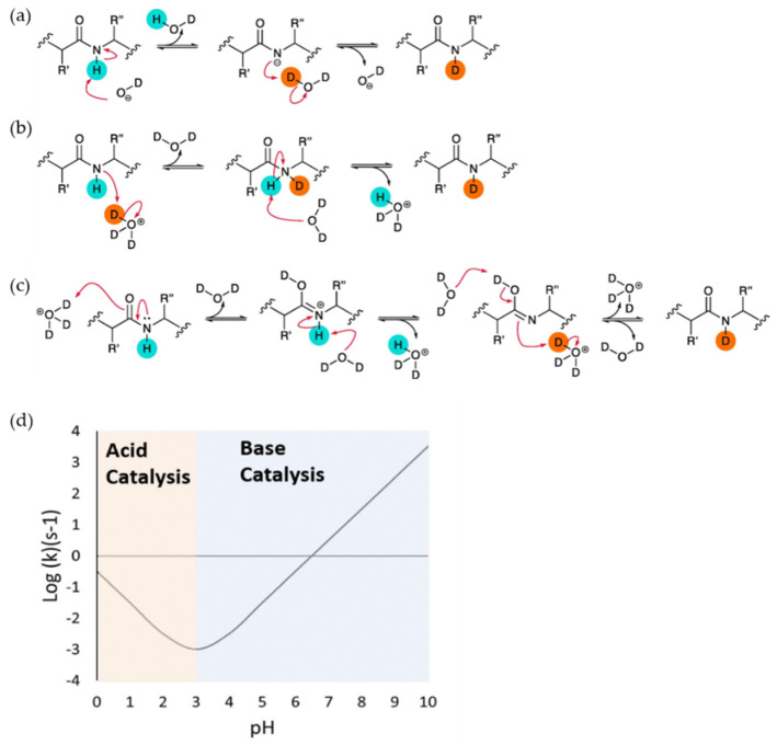
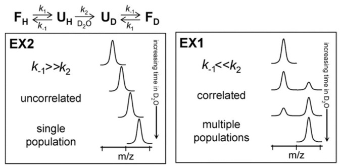
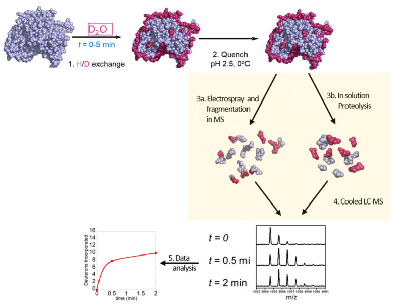
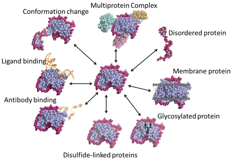
Virtually all protein functions in the cell, including pathogenic processes, require coordinated motion of atoms or domains, i.e., conformational dynamics. Understanding protein dynamics is therefore critical both for drug development and to learn about the underlying molecular causes of many diseases. Hydrogen-Deuterium Exchange Mass Spectrometry (HDX-MS) provides valuable information about protein dynamics, which is highly complementary to the static picture provided by conventional high-resolution structural tools (i.e., X-ray crystallography and structural NMR). The amount of protein required to carry out HDX-MS experiments is a fraction of the amount required by alternative biophysical techniques, which are also usually lower resolution. Use of HDX-MS is growing quickly both in industry and academia, and it has been successfully used in numerous drug and vaccine development efforts, with important roles in understanding allosteric effects and mapping binding sites.

Keywords: HDX-MS; hydrogen–deuterium exchange mass spectrometry; protein dynamics; protein folding and misfolding.


Conflict of interest statement

The authors declare no conflicts of interest.

Figures

Figure 1
Figure 1
Schematic of acid and base catalysis of peptide group protons in solution during H-D exchange and the dependence of intrinsic rate of exchange (kch) on the pH of solution. (a) Base catalysis (b) Acid catalysis through the protonation of peptide group N atom. (c) Acid catalysis through protonation of peptide group O atom. Reproduced with permission from Journal Methods@ 2018. (d) The dependence of intrinsic rate of exchange (kch) of poly-DL-alanine on the pH showing that the minimum exchange is at pH 2.5–3. Acid catalysis is dominant below the pH and base catalysis is dominant above the pH. Reproduced with permission from the journal Analyst (© 2017).
Figure 2
Figure 2
Protein unfolding kinetics and Hydrogen–Deuterium Exchange Mass Spectrometry (HDX-MS) at the top. The two possible mechanisms for hydrogen exchange in proteins, EX1 and EX2. Reproduced with permission from the Journal of the American Society for Mass Spectrometry (© 2006).
Figure 3
Figure 3
Schematic overview of top-down and bottom-up HDX-MS workflow: 1—exchange of hydrogen to deuterium for different time points (0–5 min), 2—quenching at low pH, 3a—proteolysis, 3b—electrospray and fragmentation in MS 4—separation of peptides by LC–MS and 5—data analysis.
Figure 4
Figure 4
Various applications of HDX-MS technique.
Figure 5
Figure 5
The peptide spanning 109–132 in WT and A116V moPrP displays a significant decrease in deuterium uptake in the presence of liposomes. At 30 s of deuterium labeling, there is a bimodal distribution in the presence of liposomes, compared with the unimodal distribution seen in the absence of liposomes. The controls of protonated (0% D) and deuterated (90% D) peptide fragments are also shown. Reproduced with permission from the Biophysical Society (© 2016).
Figure 6
Figure 6
(A) The deuteration map for recombinant Vif. The bars are colored according to the relative percent deuterium incorporation as indicated in the legend. Additionally indicated are the different regions of Vif and their corresponding biological functions. Representative spectra of four peptides are shown, with deuteration times included in each panel; UN represents an undeuterated protein. Reproduced with permission from Elsevier Ltd. ©2011. (B) Location of conformational changes in Elongin B and C induced by HIV-1 Vif interaction. Deuterium uptake curves for Elongin B and C peptides that displayed changes in deuterium uptake upon interaction with Vif are shown in the top panel. The solid lines represent the Elongin BC complex alone, and the dotted lines represent the Elongin BC complex bound to Vif. The changes are mapped onto the structure of the Vif140–156. Elongin BC complex. The colors in the top panel correspond to those mapped onto the structures. Areas that are grey indicate regions with no change in deuterium uptake or those which were not covered. The HIV-1 Vif peptide 140–156 is represented as the copper helix, and the remaining structurally uncharacterized regions of Vif are depicted in cartoon form. A dotted line indicates the boundary between Elongin C and Elongin B. Reproduced with permission from Elsevier Ltd. (© 2010).
Figure 7
Figure 7
(A) Difference plot generated by subtracting the absolute deuterium exchange of ligand-free ApoD from progesterone-bound ApoD. Differences > ±0.5 Da are considered significant and were found in nine peptides. Asterisks denote peptides showing bimodal exchange profiles in both the apo-form and in the presence of progesterone. (B) Absolute deuterium exchange over time of nine peptides showing significant changes upon progesterone binding. (C) Significant orthosteric changes in ApoD upon progesterone binding. (D) Zoom of the ligand binding pocket. Asn−58 is located in peptide 49–61 which shows increased deuterium exchange upon progesterone binding. Met−93 and Phe−98 are located in peptides which show decreased deuterium exchange upon progesterone binding. (E) Significant allosteric changes in ApoD upon progesterone binding. (F) Hydrogen bond network between progesterone, Tyr−46, Tyr−22, Tyr−98, and Arg−131. Reproduced with permission from The Protein Society (© 2018).
Figure 8
Figure 8
(A) Regions showing decreases in deuterium uptake upon diphtheria toxin (DTx)- monoclonal antibody (mAb) 2–25 complexation are shown in green and mapped onto the structure of DTx. The respective kinetic plots are also shown, with red and blue plots representing the uptake in the absence and presence of mAb 2–25, respectively. A reference structure with labeled domains is shown in the inset, with the dashed circle representing the HBEGF–binding pocket. (B) Regions showing decreases in deuterium uptake upon DTx-mAb 2–18 complexation are shown in red and mapped onto the structure of DTx. The respective kinetic plots are also shown, with red and blue plots representing the uptake in the absence and presence of mAb 2–18, respectively. Reproduced with permission from the American Chemical Society (© 2018).
Figure 9
Figure 9
(A) Subunit architecture of FOF1 ATP synthase from E. coli. The bottom panel depicts membrane vesicle-bound FOF1 under different experimental conditions. (B) HDX levels of IADP for an HDX period of 45 min. (C) Deuteration difference map of WPMF vs. IADP. (D) Deuteration difference map of WFCCP vs. IADP. Regions that were not covered are shown in dark gray. Colors represent deuteration percentages or HDX difference (%) as indicated in the legends. Reproduced with permission from the National Academy of Sciences (© 2016).
Figure 10
Figure 10
(A) Most representative structure for the native ensemble, which exhibits a global fold with sequestration of the hexapeptides. The structure is colored based on protection factor, as indicated in the legend. (B) Most representative structure for the hyperphosphorylated ensemble showing release of the N- and C-termini containing the microtubule-binding region, full exposure of H2 and a few regions of residual structure, including H1. Reproduced with permission from PLoS ONE (© 2015).
Figure 11
Figure 11
Cartoon illustration of β2-Adrenergic G-protein coupled receptor (GPCR) (β2AR) in (A) a lipid bicelle (3:1 DMPC:CHAPSO) and (B) dodecyl maltoside (DDM). (C) The sequence coverage of β2AR prepared in bicelles and (D) DDM. Blue bars represent peptic peptides identified from four independent experiments. Reproduced with permission from the American Chemical Society (© 2015).
Figure 12
Figure 12
(A) HDX-MS workflow with in-solution deglycosylation at low pH using PNGase. HDX-MS sequence coverage of Trastuzumab (B) heavy chain and (C) light chain using the optimized workflow. The glycosylated residue N301 and the deamidated deglycosylated residue D301 are shown in blue and red, respectively, along with the detected N-linked glycopeptides. Reproduced with permission from the American Chemical Society (© 2016).

Similar articles

Cited by

References

    1. Soto C. Unfolding the role of protein misfolding in neurodegenerative diseases. Nat. Rev. Neurosci. 2003;4:49–60. doi: 10.1038/nrn1007. - DOI - PubMed
    1. Zhao S., Wu B., Stevens R.C. Advancing Chemokine GPCR Structure Based Drug Discovery. Structure. 2019;27:405–408. doi: 10.1016/j.str.2019.02.004. - DOI - PubMed
    1. Deng B., Lento C., Wilson D.J. Hydrogen deuterium exchange mass spectrometry in biopharmaceutical discovery and development — A review. Anal Chim. Acta. 2016;940:8–20. doi: 10.1016/j.aca.2016.08.006. - DOI - PubMed
    1. Berkowitz S.A., Engen J.R., Mazzeo J.R., Jones G.B. Analytical tools for characterizing biopharmaceuticals and the implications for biosimilars. Nat. Rev. Drug Discov. 2012;11:527–540. doi: 10.1038/nrd3746. - DOI - PMC - PubMed
    1. Beck A., Wagner-Rousset E., Ayoub D., Dorsselaer V.A., Sanglier-Cianférani S. Characterization of Therapeutic Antibodies and Related Products. Anal Chem. 2013;85:715–736. doi: 10.1021/ac3032355. - DOI - PubMed

LinkOut - more resources