. 2020 Sep 1;205(5):1323-1330.
doi: 10.4049/jimmunol.2000144. Epub 2020 Jul 24.

Killer Cell Immunoglobulin-like Receptor Variants Are Associated with Protection from Symptoms Associated with More Severe Course in Parkinson Disease

Affiliations

Killer Cell Immunoglobulin-like Receptor Variants Are Associated with Protection from Symptoms Associated with More Severe Course in Parkinson Disease

Kirsten M Anderson et al. J Immunol. .

Abstract

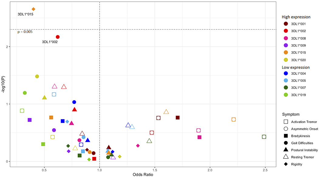
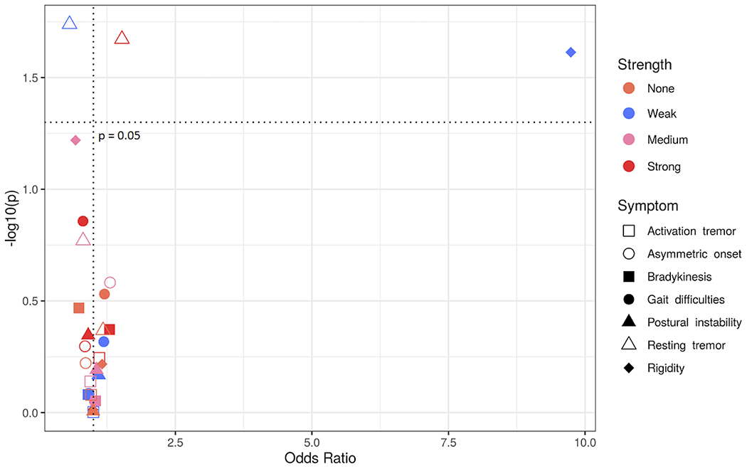
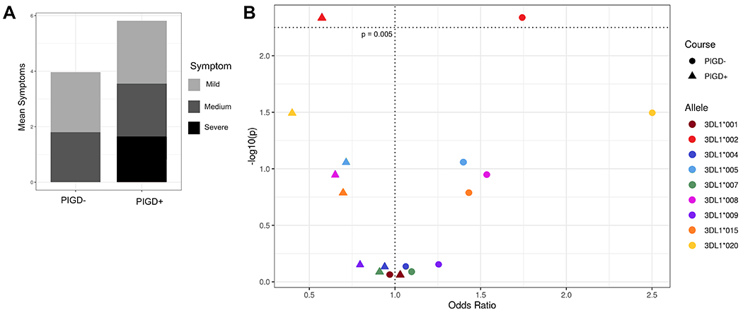
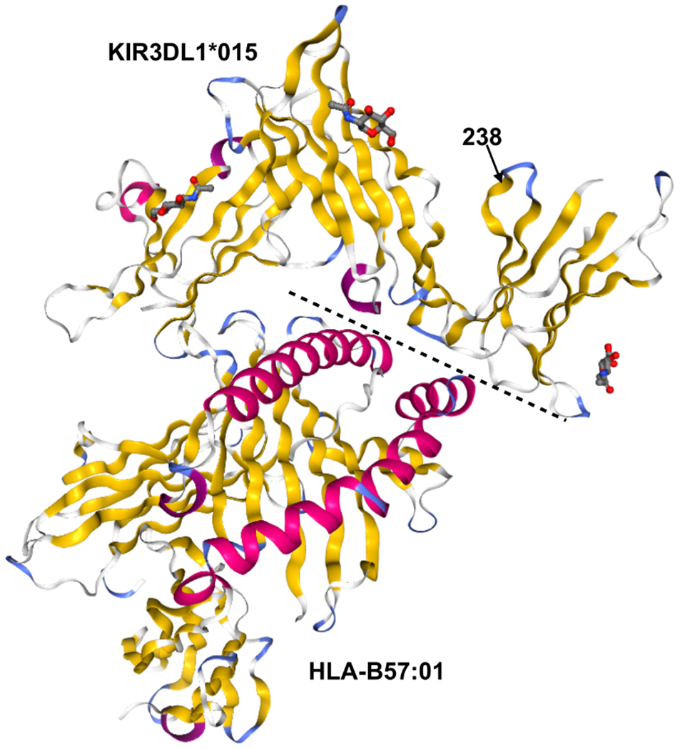
Immune dysfunction plays a role in the development of Parkinson disease (PD). NK cells regulate immune functions and are modulated by killer cell immunoglobulin-like receptors (KIR). KIR are expressed on the surface of NK cells and interact with HLA class I ligands on the surface of all nucleated cells. We investigated KIR-allelic polymorphism to interrogate the role of NK cells in PD. We sequenced KIR genes from 1314 PD patients and 1978 controls using next-generation methods and identified KIR genotypes using custom bioinformatics. We examined associations of KIR with PD susceptibility and disease features, including age at disease onset and clinical symptoms. We identified two KIR3DL1 alleles encoding highly expressed inhibitory receptors associated with protection from PD clinical features in the presence of their cognate ligand: KIR3DL1*015/HLA-Bw4 from rigidity (p c = 0.02, odds ratio [OR] = 0.39, 95% confidence interval [CI] 0.23-0.69) and KIR3DL1*002/HLA-Bw4i from gait difficulties (p c = 0.05, OR = 0.62, 95% CI 0.44-0.88), as well as composite symptoms associated with more severe disease. We also developed a KIR3DL1/HLA interaction strength metric and found that weak KIR3DL1/HLA interactions were associated with rigidity (pc = 0.05, OR = 9.73, 95% CI 2.13-172.5). Highly expressed KIR3DL1 variants protect against more debilitating symptoms of PD, strongly implying a role of NK cells in PD progression and manifestation.


Conflict of interest statement

Conflict of Interest Statement: The authors have no conflicts of interest to disclose.

Figures

Figure 1.
Figure 1.. KIR3DL1 alleles associated with protection from PD symptoms.
All KIR3DL1 alleles were analyzed in combination with HLA-Bw4 ligands. Odds ratios (x – axis) by -log10(p-value) (y – axis). Each dot represents the odds ratio and corresponding -log10(p-value) for a KIR allele/symptom pair. PD symptoms are indicated by shapes and KIR3DL1 alleles are indicated by color. The horizontal dotted line indicates the significant corrected p-value.
Figure 2.
Figure 2.. Strong interactions between KIR3DL1 and HLA-Bw4 alleles are associated with clinical features of PD.
Odds ratios (x – axis) by -log10(p-value) (y – axis). Each dot represents the odds ratio and corresponding -log10(p-value) for a KIR interaction strength/symptom pair. Different PD symptoms are indicated by shapes and KIR3DL1 interaction strengths are indicated by color.
Figure 3.
Figure 3.. Stratification of PD patients by symptom type.
A) Mean number of symptoms (y – axis) per cluster (x – axis). Symptoms are broken down into three categories where red indicates the most debilitating (postural instability and gait difficulty), coral the moderate symptoms (rigidity and bradykinesis), and light pink the mild symptoms associated with less severe disease (resting and activation tremors and asymmetric onset). B) KIR3DL1 allele-HLA-Bw4 associations with disease group. Odds ratio (x – axis) by -log10(p-value) (y – axis). Each dot represents a KIR3DL1 allele-HLA ligand pair, the different shapes indicate PIGD- (circle) or PIGD+ (triangle) disease. The horizontal dotted line indicates the value for a significant corrected p-value.
Figure 4.
Figure 4.. KIR3DL1*015 and KIR3DL1*002 Differ by A Single Amino Acid at Position 238.
Crystal structures of KIR3DL1*015 (top) complexed with its ligand HLA-B*57:01 (bottom). Secondary structures are shown by color: alpha helices (pink) and beta sheets (yellow). The dashed line indicates the binding interface between KIR3DL1*015 and HLA-B*57:01. Position 238 is indicated by an arrow. Protein Data Bank accession no. 5B39 (41).
Figure 5.
Figure 5.. Molecular dynamics simulations of KIR3DL1*015 and KIR3DL1*002.
A) KIR3DL1 is composed of three extracellular domains (D0-D2). The HLA binding site (purple) is located at the hinge between D1 and D2. Residue 238 (yellow) is in the D2 domain facing towards D1 serving as an anchorage point. The G to R substitution in 3DL1*002 positions D2 closer to D1 allowing more contacts between surface residues of D1 and D2. B) This led to an increased D1-D2 interaction energy by 50 kcal/mol in 3DL1*002 compared to 3DL1*015. C) The association between residue 238 and D1 was also 10-fold higher in 3DL1*002 as opposed to 3DL1*015. D) Residue cross correlation (RCC) map depicts pairwise correlations between residue 238 and the HLA binding site, mapped on residues P163, M165, L166, A167, P199, Y200, E201, S227, S228, D230, F276, R277, H278, S279, Y281, and E282. The negative correlation between G238 and residues E201, S227, S228, and D230 in 3DL1*015 was eliminated upon G238R substitution in 3DL1*002 as shown by the dashed boxes. On the contrary, the positive correlation between HLA binding residues is enhanced in 3DL1*002 (solid boxes).

Similar articles

Cited by

References

    1. Polymeropoulos MH, Higgins JJ, Golbe LI, Johnson WG, Ide SE, Di Iorio G, Sanges G, Stenroos ES, Pho LT, Schaffer AA, Lazzarini AM, Nussbaum RL, and Duvoisin RC. 1996. Mapping of a gene for Parkinson’s disease to chromosome 4q21-q23. Science 274: 1197–1199. - PubMed
    1. Nussbaum RL, and Polymeropoulos MH. 1997. Genetics of Parkinson’s disease. Hum Mol Genet 6: 1687–1691. - PubMed
    1. Gorell JM, Johnson CC, Rybicki BA, Peterson EL, and Richardson RJ. 1998. The risk of Parkinson’s disease with exposure to pesticides, farming, well water, and rural living. Neurology 50: 1346–1350. - PubMed
    1. Hernan MA, Takkouche B, Caamano-Isorna F, and Gestal-Otero JJ. 2002. A meta-analysis of coffee drinking, cigarette smoking, and the risk of Parkinson’s disease. Ann Neurol 52: 276–284. - PubMed
    1. Hollenbach JA, Norman PJ, Creary LE, Damotte V, Montero-Martin G, Caillier S, Anderson KM, Misra MK, Nemat-Gorgani N, Osoegawa K, Santaniello A, Renschen A, Marin WM, Dandekar R, Parham P, Tanner CM, Hauser SL, Fernandez-Vina M, and Oksenberg JR. 2019. A specific amino acid motif of HLA-DRB1 mediates risk and interacts with smoking history in Parkinson’s disease. Proc Natl Acad Sci U S A 116: 7419–7424. - PMC - PubMed

Publication types