Review
. 2021 Apr;105(4):454-467.
doi: 10.1136/bjophthalmol-2020-316149. Epub 2020 Jul 24.

Evolution of therapies for the corneal endothelium: past, present and future approaches

Affiliations
Review

Evolution of therapies for the corneal endothelium: past, present and future approaches

Hon Shing Ong et al. Br J Ophthalmol. 2021 Apr.

Erratum in

Abstract

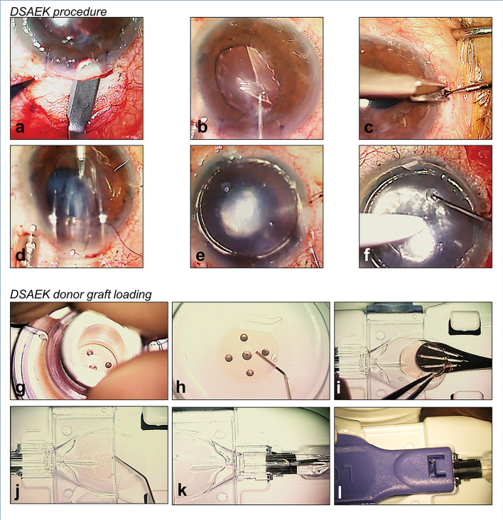
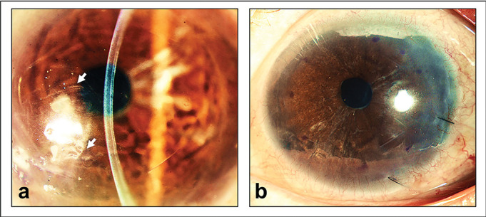
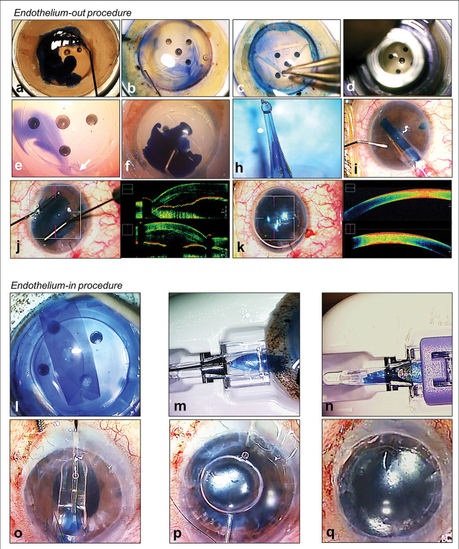
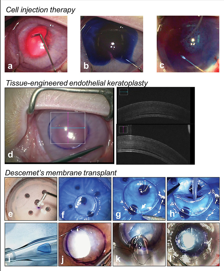
Corneal endothelial diseases are leading indications for corneal transplantations. With significant advancement in medical science and surgical techniques, corneal transplant surgeries are now increasingly effective at restoring vision in patients with corneal diseases. In the last 15 years, the introduction of endothelial keratoplasty (EK) procedures, where diseased corneal endothelium (CE) are selectively replaced, has significantly transformed the field of corneal transplantation. Compared to traditional penetrating keratoplasty, EK procedures, namely Descemet's stripping automated endothelial keratoplasty (DSAEK) and Descemet membrane endothelial keratoplasty (DMEK), offer faster visual recovery, lower immunological rejection rates, and improved graft survival. Although these modern techniques can achieve high success, there are fundamental impediments to conventional transplantations. A lack of suitable donor corneas worldwide restricts the number of transplants that can be performed. Other barriers include the need for specialized expertise, high cost, and risks of graft rejection or failure. Research is underway to develop alternative treatments for corneal endothelial diseases, which are less dependent on the availability of allogeneic tissues - regenerative medicine and cell-based therapies. In this review, an overview of past and present transplantation procedures used to treat corneal endothelial diseases are described. Potential novel therapies that may be translated into clinical practice will also be presented.

Keywords: Angiogenesis; Choroid; Fuchs’ endothelial dystrophy; Genetics; Imaging; Infection; Inflammation; Ocular surface; Stem Cells; Treatment Lasers; bullous keratopathy; cell therapy; cornea; corneal endothelium; corneal transplantation; eye (tissue) banking; keratoplasty; regenerative medicine.


Conflict of interest statement

Competing interests: JSM holds a patent on the EndoGlide and receive royalties. The other authors have no other relevant affiliations or financial involvement with any organisation or entity with a financial interest in or financial conflict with the subject matter or materials discussed in the manuscript apart from those disclosed.

Figures

Figure 1
Figure 1
Authors’ preferred surgical technique of Descemet’s stripping automated endothelial keratoplasty (DSAEK) with the EndoGlide (Network Medical Products, Yorkshire, UK). (A) 4.5 mm scleral-tunnelled surgical wound; (B) descemetorhexis; (C) inferior peripheral iridectomy; (D) pre-cut DSAEK lenticule inserted into the anterior chamber via a pull-through technique; (E) injection of air for graft attachment; (F) opening of venting incisions to release fluid from the graft–host interface; (G) trephination of pre-cut graft; (H) fluid separation of DSAEK lenticule from anterior stromal cap; (I) DSAEK lenticule transferred to the EndoGlide; (J) ocular viscoelastic device coating to protect donor endothelial cells; (K) DSAEK lenticule is pulled into the EndoGlide using a customised micro-forceps; and (L) clip secured creating a ‘closed system’ during DSAEK graft insertion.
Figure 2
Figure 2
Optical quality degradation following Descemet’s stripping automated endothelial keratoplasty (DSAEK). (A) Patient who received a DSAEK graft showing folds visible in the interface (arrows) between graft and recipient stroma resulting in visual symptoms; (B) the same patient who had a DSAEK graft exchanged with a Descemet’s membrane endothelial keratoplasty (DMEK) graft showing the resolution of folds and corresponding improvement in optical quality.
Figure 3
Figure 3
Authors’ preferred surgical techniques of Descemet’s membrane endothelial keratoplasty (DMEK). (A–K) Donor preparation using submerged cornea using backgrounds away (SCUBA) technique and ‘endothelium-out’ surgical technique of DMEK. (A) Visionblue (D.O.R.C., Zuidland, The Netherlands) to enhance Descemet’s membrane (DM) visualisation; (B) corneal tissue scored in the periphery; (C) DM is peeled from periphery; (D) DMEK graft is trephined; (E) arrow showing surgically cut orientation mark (asymmetrical triangle); (F) Membrane Blue Dual (D.O.R.C., Zuidland, The Netherlands) applied to DMEK graft; (H) loading of graft into glass-injector; (I) intracameral injection of DMEK graft via a corneal surgical wound; (J) graft is unfolded using controlled taps over host cornea; (K) injection of air/gas for graft attachment; (L–Q) Surgical technique of ‘endothelium-in’ DMEK using a DMEK EndoGlide (Network Medical Products, Yorkshire, UK). (L) creating an ‘endothelium-in’ graft trifold; (M) donor loading into the DMEK EndoGlide using a pull-through technique; (N) clip secured creating a ‘closed system’ during DMEK graft insertion; (O) graft inserted into the anterior chamber by a pull-through technique; (P) air is injected whilst holding the donor to maintain orientation; (Q) intracameral full air/gas fill for graft attachment.
Figure 4
Figure 4
Strategies to improve success of Descemet’s membrane endothelial keratoplasty (DMEK). (A) Air used to separate and detach Descemet’s membrane from stroma for donor harvest; (B) eye bank pre-stripped DMEK graft; note the orientation S-stamp and mark on scleral rim indicating the unstripped area of the graft (arrow); (C–F) example of a tight scroll and use of air bubbles to assist the surgeon in opening up the graft (D, E) before full air tamponade (F).
Figure 5
Figure 5
Novel therapies for the treatment of corneal endothelial diseases. (A–C) Cell injection therapy performed in a rabbit model of bullous keratopathy; silicone-tipped cannula is used to remove native corneal endothelial cells guided by trypan blue (A, B) followed by injection of cultured human corneal endothelial cells (C); (D) tissue-engineered endothelial keratoplasty performed in a rabbit model; (E–L) technique of the Descemet’s membrane transplant (DMT); (E) removal of donor corneal endothelial cells by Descemet’s membrane (DM) scrapping with a silicone tip cannula (SP-125053, ASICO, USA); (F) trephination of DMT graft; note posterior surface of graft is stained blue with Visionblue (D.O.R.C, Zuidland, The Netherlands), showing its aceullarity; (G) orientation mark created (asymmetrical scalene triangle); (H) acellular DM disc is the loaded into a glass injector; (J) surgical marking on host cornea (4–5 mm) to indicate the central diseased area of DM stripping; (K) intracameral injection of DMT graft via a corneal surgical wound; (L) intracameral full air/gas fill for graft attachment.

References

    1. Bonanno JA. Molecular mechanisms underlying the corneal endothelial pump. Exp Eye Res 2012;95:2–7. 10.1016/j.exer.2011.06.004 - DOI - PMC - PubMed
    1. Edelhauser HF. The balance between corneal transparency and edema: the proctor lecture. Invest Ophthalmol Vis Sci 2006;47:1754–67. 10.1167/iovs.05-1139 - DOI - PubMed
    1. Bahn CF, Glassman RM, MacCallum DK, et al. Postnatal development of corneal endothelium. Invest Ophthalmol Vis Sci 1986;27:44–51. - PubMed
    1. Bourne WM. Biology of the corneal endothelium in health and disease. Eye (Lond) 2003;17:912–8. 10.1038/sj.eye.6700559 - DOI - PubMed
    1. Nucci P, Brancato R, Mets MB, et al. Normal endothelial cell density range in childhood. Arch Ophthalmol 1990;108:247–8. 10.1001/archopht.1990.01070040099039 - DOI - PubMed