Review
. 2021 Feb;35(2):362-382.
doi: 10.1038/s41433-020-1096-5. Epub 2020 Jul 24.

Refractive surgery beyond 2020

Affiliations
Review

Refractive surgery beyond 2020

Marcus Ang et al. Eye (Lond). 2021 Feb.

Abstract

Refractive surgery refers to any procedure that corrects or minimizes refractive errors. Today, refractive surgery has evolved beyond the traditional laser refractive surgery, embodied by the popular laser in situ keratomileusis or 'LASIK'. New keratorefractive techniques such as small incision lenticule extraction (SMILE) avoids corneal flap creation and uses a single laser device, while advances in surface ablation techniques have seen a resurgence in its popularity. Presbyopic treatment options have also expanded to include new ablation profiles, intracorneal implants, and phakic intraocular implants. With the improved safety and efficacy of refractive lens exchange, a wider variety of intraocular lens implants with advanced optics provide more options for refractive correction in carefully selected patients. In this review, we also discuss possible developments in refractive surgery beyond 2020, such as preoperative evaluation of refractive patients using machine learning and artificial intelligence, potential use of stromal lenticules harvested from SMILE for presbyopic treatments, and various advances in intraocular lens implants that may provide a closer to 'physiological correction' of refractive errors.

摘要: 屈光手术是指矫正或减少屈光不正度数的任何手术。如今, 屈光手术已经超越了传统的手术方式, 以流行的激光原位角膜磨镶术或”LASIK”为代表(laser in-situ keratomileusis, LASIK)。新的角膜屈光技术如小切口角膜基质透镜取出术 (small incision lenticule extraction, SMILE) 避免了角膜瓣的产生和使用单一激光设备, 另外, 表面消融技术的进步使其的流行度回升。老花眼治疗方案也已扩展到涉及新的消融方式, 角膜内植入物和人工晶状体植入物等方面。随着屈光镜片更换的安全性和功效的提高, 具有更先进光学技术的人工晶状体植入物为精心挑选的患者提供了更多屈光矫正手术的选择。在这篇综述中, 讨论了2020年以后屈光手术可能的发展, 例如机器学习和人工智能用于屈光手术患者的术前评估, 从SMILE中获得的角膜基质透镜在老视治疗中的潜在应用, 以及人工晶状体植入物的各种进步可以提供更接近屈光不正的“生理性的矫正”。.


Conflict of interest statement

MA has no financial conflicts of interests to declare. DZR is a consultant for Carl Zeiss Meditec AG (Jena, Germany) DZR acknowledges a financial interest in Artemis Insight 100 VHF digital ultrasound (ArcScan Inc., Golden, CO)

Figures

Fig. 1
Fig. 1. High-resolution swept-source optical coherence tomography imaging of the anterior segment of a pseudophakic eye.
High-resolution swept-source OCT imaging of the anterior segment of a pseudophakic eye in which after piggy-back implantation of a sulcus lens to fix a refractive surprise after uneventful cataract surgery (ANTERION, Heidelberg Engineering GmbH, Heidelberg, Germany).
Fig. 2
Fig. 2. Precision high-frequency ultrasound device (50 MHz) imaging of the cornea.
Ultrasound imaging can accurately image behind the iris allowing for improved ICL sizing. In addition to measurements of the anatomic structures comprising the anterior of the eye such as anterior chamber, the instrument can delineate the corneal epithelial layer thickness (ArcScan Insight 100, ArcScan Inc, Golden, USA).
Fig. 3
Fig. 3. Average lens density (ALD) quantification with swept-source optical coherence tomography (SS-OCT).
Six radial B-scans including the lens were acquired three times at each meridian (0, 30, 60, 90, 120, and 150 degrees). An algorithm measured the density of the region of interest on a scale of 0–255 pixel intensity units. In a recent study, an SS-OCT ALD measurement of 73.8 pixel units or greater strongly suggested the presence of cataract, with a sensitivity of 96.2% and a specificity of 91.3%.
Fig. 4
Fig. 4. Central flap thickness reproducibility comparing microkeratome and femtosecond laser.
Graph showing the central flap thickness reproducibility for all studies published between 1998 and 2014, grouped by mechanical microkeratome (blue) and femtosecond laser (red).
Fig. 5
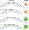
Fig. 5. Series of diagrams showing the femtosecond cutting sequence for a SMILE procedure.
(1) lenticule interface from out-to-in, (2) lenticule side cut, (3) cap interface from in-to-out, and (4) the small incision(s). Reprinted with permission from Reinstein et al. [117].
Fig. 6
Fig. 6. Series of images showing the standard surgical technique for SMILE.
Reprinted with permission from Reinstein et al. [117].
Fig. 7
Fig. 7. Corneal stroma enhancement with a decellularized corneal stroma lenticule in a patient with advanced keratoconus.
Slit-lamp pictures 1 week (top-left) and 3 months after surgery (top-right; note the complete transparency recovery). Corneal OCT image (down): the implanted lenticule is easily identified 6 months after surgery (white arrows).
Fig. 8
Fig. 8. Slit-lamp photograph of an eye with intracorneal implants.
Intracorneal ring implanted in a patient with keratoconus.
Fig. 9
Fig. 9. Raindrop inlay.
Slit lamp (left; white arrows point the edges of the inlay) and anterior segment optical coherence tomography (OCT) pictures (right). Observe how the thin inlay is seen in the OCT image and it appears surrounded by a mild stromal haze (yellow dashed line).
Fig. 10
Fig. 10. Kamra inlay.
Slit-lamp pictures 3 months (left; observe the peripheral microperforations to allow corneal nutrition) and 3 years after implantation (middle). Note the progressive moderate haze associated with visual loss that justified inlay explantation, remaining a donut-shape central corneal scar still visible 4 years after inlay removal (right).
Fig. 11
Fig. 11. Anterior chamber iris-claw intraocular lenses.
Left: rigid polymethyl-methacrylate (PMMA) lens. Right: foldable lens made of polysiloxane. Both from Ophtec BV, the Netherlands and J&J, USA.
Fig. 12
Fig. 12. Example of implantable collamer lens (ICL) in situ.
The central port allows aqueous flow from the posterior chamber to the anterior chamber in order to maintain the normal physiology of the anterior segment of the eye. The EVO+ Visian ICL (STAAR Surgical, USA) introduced in 2016 now was an extended optical zone.
Fig. 13
Fig. 13. Example of multifocal intraocular lens implants recently available.
Left: trifocal intraocular lens implant. Right: hybrid design extended depth of focus (EDOF) intraocular lens implant.
Fig. 14
Fig. 14. Photographs of a small aperture intraocular lens implant before and after implantation in situ.
Example of small aperture intraocular lens implant, which utilizes the pinhole principle to increase the depth of focus to about 3D.

Comment in

References

    1. Kim TI, Alio Del Barrio JL, Wilkins M, Cochener B, Ang M. Refractive surgery. Lancet. 2019;393:2085–98. - PubMed
    1. Sugar A, Hood CT, Mian SI. Patient-reported outcomes following LASIK: quality of Life in the PROWL Studies. JAMA. 2017;317:204–5. - PubMed
    1. Sugar A, Rapuano CJ, Culbertson WW, Huang D, Varley GA, Agapitos PJ, et al. Laser in situ keratomileusis for myopia and astigmatism: safety and efficacy: a report by the American Academy of Ophthalmology. Ophthalmology. 2002;109:175–87. - PubMed
    1. Sandoval HP, Donnenfeld ED, Kohnen T, Lindstrom RL, Potvin R, Tremblay DM, et al. Modern laser in situ keratomileusis outcomes. J Cataract Refract Surg. 2016;42:1224–34. - PubMed
    1. Eydelman M, Hilmantel G, Tarver ME, Hofmeister EM, May J, Hammel K, et al. Symptoms and satisfaction of patients in the patient-reported outcomes with laser in situ keratomileusis (PROWL) studies. JAMA Ophthalmol. 2017;135:13–22. - PubMed