. 2020 Sep;140(3):359-375.
doi: 10.1007/s00401-020-02189-9. Epub 2020 Jul 24.

Lesion stage-dependent causes for impaired remyelination in MS

Affiliations

Lesion stage-dependent causes for impaired remyelination in MS

Katharina Heß et al. Acta Neuropathol. 2020 Sep.

Abstract

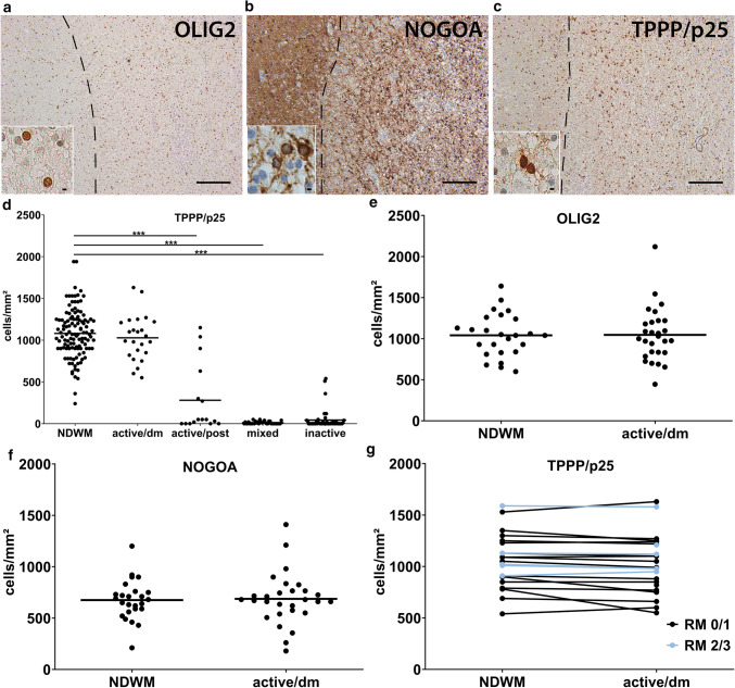
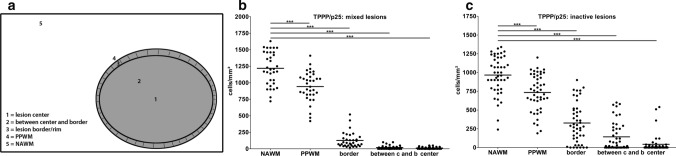
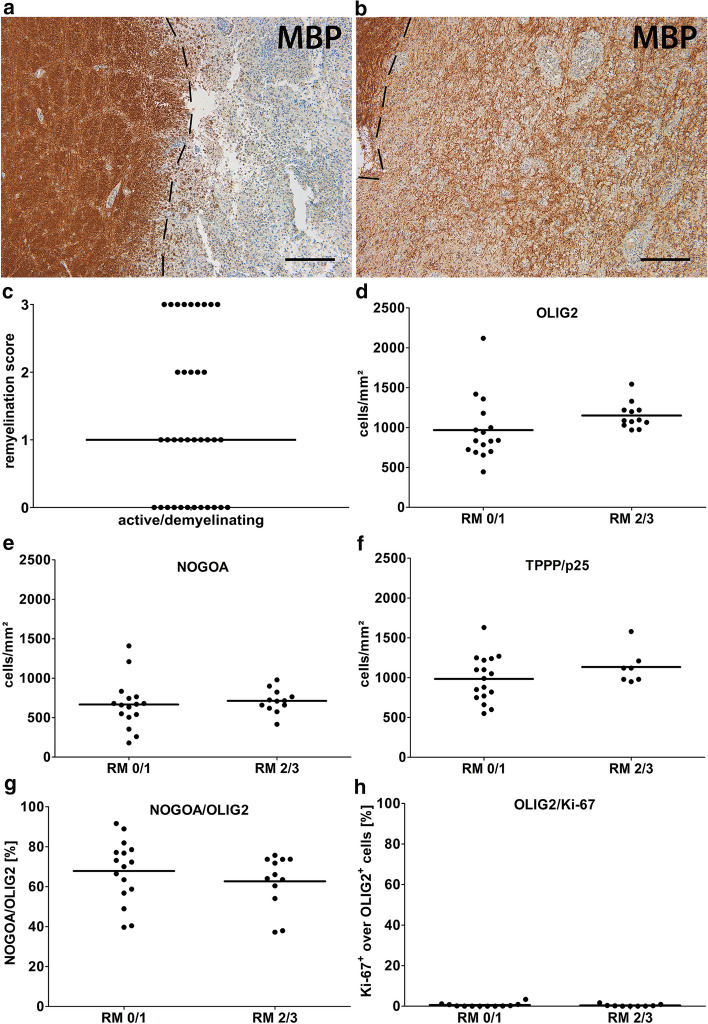
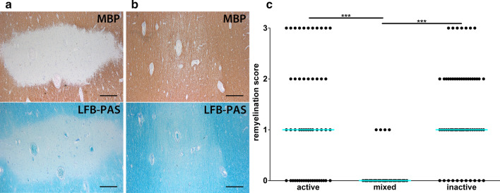
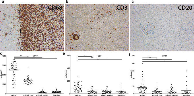
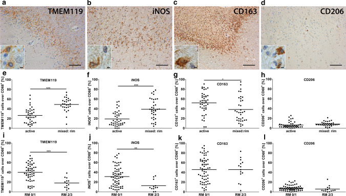
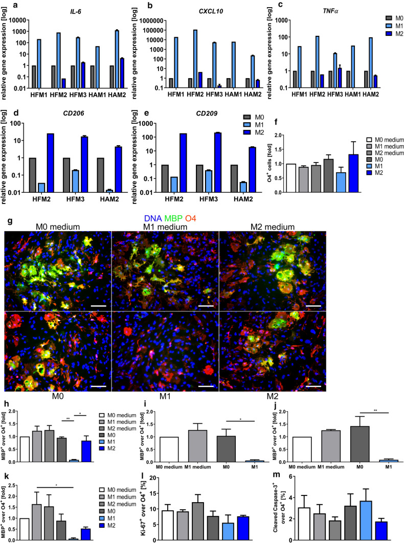
Multiple sclerosis (MS) is the most frequent demyelinating disease and a leading cause for disability in young adults. Despite significant advances in immunotherapies in recent years, disease progression still cannot be prevented. Remyelination, meaning the formation of new myelin sheaths after a demyelinating event, can fail in MS lesions. Impaired differentiation of progenitor cells into myelinating oligodendrocytes may contribute to remyelination failure and, therefore, the development of pharmacological approaches which promote oligodendroglial differentiation and by that remyelination, represents a promising new treatment approach. However, this generally accepted concept has been challenged recently. To further understand mechanisms contributing to remyelination failure in MS, we combined detailed histological analyses assessing oligodendroglial cell numbers, presence of remyelination as well as the inflammatory environment in different MS lesion types in white matter with in vitro experiments using induced-pluripotent stem cell (iPSC)-derived oligodendrocytes (hiOL) and supernatants from polarized human microglia. Our findings suggest that there are multiple reasons for remyelination failure in MS which are dependent on lesion stage. These include lack of myelin sheath formation despite the presence of mature oligodendrocytes in a subset of active lesions as well as oligodendroglial loss and a hostile tissue environment in mixed active/inactive lesions. Therefore, we conclude that better in vivo and in vitro models which mimic the pathological hallmarks of the different MS lesion types are required for the successful development of remyelination promoting drugs.

Keywords: Microglia; Multiple sclerosis; Oligodendrocytes; Remyelination.


Conflict of interest statement

T.K. has a pending patent application for the generation of human oligodendrocytes.

Figures

Fig. 1
Fig. 1
Preservation of oligodendrocytes in active/demyelinating lesions. ac OLIG2+, NOGOA+ and TPPP/p25+ oligodendrocytes were identified using IHC. Inserts show oligodendrocytes in higher magnification. d Quantification of oligodendrocytes in different lesion types and NDWM using TPPP/p25. e, f Quantification of OLIG2+ and NOGOA+ oligodendrocytes demonstrate comparable cell numbers in active/demyelinating as well as in NDWM. g When comparing tissue samples containing NDWM and active/demyelinating lesions, no significant differences in numbers of TPPP/p25+ oligodendrocytes were observed. Lesions with marked remyelination are indicated in blue, lesions with limited remyelination in black. Scale bars in a to c: 200 µm, scale bars in the inserts in a to c 6.25 µm. OLIG2 oligodendrocyte transcription factor, TPPP/p25 tubulin polymerization promoting protein, NDWM non-demyelinated white matter, active/dm active/demyelinating lesions, active/post active/post-demyelinating lesions, mixed mixed active/inactive lesions, inactive inactive lesions, RM 0/1 remyelination score 0 or 1, RM 2/3 remyelination score 2 or 3
Fig. 2
Fig. 2
Loss of oligodendrocytes in mixed and inactive lesions. a Schematic drawing indicating the different areas in which oligodendroglial cell numbers were quantified. b, c Immunohistochemistry for TPPP/p25 revealed continuous decrease in TPPP/p25+ oligodendrocytes from NAWM to the lesion center in mixed and inactive lesions. TPPP/p25 tubulin polymerization promoting protein, PPWM periplaque white matter, NAWM normal appearing white matter, border lesion border/rim, between c and b between center and border of lesion, center lesion center
Fig. 3
Fig. 3
No differences in oligodendroglial cell numbers in active/demyelinating lesions with and without marked remyelination. a, b A lesion with no remyelination (score 0) is shown in a, whereas in b, a lesion with marked remyelination (score 3) is displayed (IHC for MBP). c In 14 out of 36 (= 39%) of the active/demyelinating lesions, we observed marked remyelination (score 2/3). df Quantification of OLIG2+, NOGOA+ and TPPP/p25+ cells in active/demyelinating lesions with (score 2/3) and without (score 0/1) marked remyelination. g No significant difference in the ratio of NOGOA+ cells over OLIG2+ in lesions with and without marked remyelination. h Double IHC for Ki-67 and OLIG2 demonstrated that only few OLIG2+ cells express Ki-67. Scale bars in a and b: 200 µm. MBP myelin basic protein, OLIG2 oligodendrocyte transcription factor 2, TPPP/p25 tubulin polymerization promoting protein, active/demyelinating active/demyelinating lesions, RM 0/1 remyelination score 0 or 1, RM 2/3 remyelination score 2 or 3
Fig. 4
Fig. 4
Almost complete lack of remyelination in mixed lesions. a, b Pictures display an inactive lesion with no remyelination (score 0) (a), whereas b shows a completely remyelinated shadow plaque (score 3). Upper panels in a and b show IHC for MBP, lower panels in a and b display LFB-PAS staining. c Semiquantitative analysis of remyelination reveals an almost complete lack of remyelination in mixed lesions (c). Scale bars in a and b: 500 µm. MBP myelin basic protein, LFB-PAS luxol fast blue–periodic acid Schiff; active active lesions, mixed mixed active/inactive lesions, inactive inactive lesions
Fig. 5
Fig. 5
Inflammatory environment in different lesion types. ac IHC demonstrates numerous CD68+ myeloid cells, fewer CD3+ T cells and few CD20+ B cells in an active lesion. All pictures are taken from the same active lesion. df Quantification of CD68+, CD3+ and CD20+ cells in different lesion types. Please note the different y-axes. Scale bar in a: 200 µm, scale bars in b and c: 100 µm. CD3/20/68 cluster of differentiation 3/20/68, active active lesions, mixed: rim  rim of mixed active/inactive lesions, mixed: center center of mixed active/inactive lesions, inactive inactive lesions
Fig. 6
Fig. 6
Relative increase in the numbers of TMEM119+ and iNOS+ myeloid cells in the rim of mixed lesions. ad IHC for TMEM119, iNOS, CD163 and CD206 was performed. Inserts show labelled cells in higher magnification. eg Quantification demonstrated a significant relative increase in the ratio of TMEM119+/CD68+ and iNOS+/CD68+ myeloid cells and a relative decrease of CD163+/CD68+ myeloid cells in the rim of mixed lesions compared to active lesions. h No differences in the number of CD206+ over CD68+ cells were observed between active lesions and the rim of mixed lesions. i, j Comparably, we observed a relative increase in the ratio of TMEM119+/CD68+ and iNOS+/CD68+ in lesions with limited (score 0/1) compared to lesions with marked remyelination (score 2/3). k, l No differences were found between lesions with and without marked remyelination with respect to the ratio of CD163+/CD68+ and CD203+/CD68+ cells. Scale bars in a, b and d: 100 µm, scale bar in c: 200 µm, scale bar in insert in ad: 6.25 µm. TMEM119 transmembrane protein 119, iNOS inducible nitric oxide synthase, CD68/163/206 cluster of differentiation 68/163/206, active active lesions, mixed: rim rim of mixed active/inactive lesions, RM 0/1 remyelination score 0 or 1, RM 2/3 remyelination score 2 or 3
Fig. 7
Fig. 7
Supernatants of primary human M1, but not M2 or M0 polarized microglia inhibit the terminal differentiation of hiOL. Every experiment analyzing the effect of microglia supernatants on hiOL was performed with hiOL derived from three different donors. ae To confirm successful polarization into M1 and M2 microglia, qRT-PCR was performed for IL-6, CXCL10 and TNFα (M1) and CD206 and CD209 (M2). f Differentiation of hiOL in the presence of supernatants from M0, M1 or M2 polarized microglia from one fetal donor (HFM2) from day 4 to 21 resulted in no significant differences in the percentage of O4+ hiOL when comparing the effect of different supernatants with appropriate controls. g, h Culturing O4 sorted hiOL in the presence of supernatants from M1, but not M0 or M2 polarized microglia from one fetal donor (HFM3) from day 21 to 35 impaired significantly the differentiation of O4+ hiOL into MBP+ mature oligodendrocytes. ik This was confirmed using supernatants from M1 and M0 polarized microglia from another fetal (HFM1) and one adult donor (HAM1) as well as supernatants from M0, M1 and M2 polarized microglia from a second adult donor (HAM2). l Analysis of percentages of actively dividing hiOL (Ki-67+ over O4+ cells) in the presence of supernatants from M0, M1 or M2 polarized microglia from one fetal donor (HFM2) from day 21 to 35 of differentiation did not reveal any significant differences. m Percentages of apoptotic hiOL (cleaved caspase 3+ over O4+ cells) in the presence of supernatants from M0, M1 or M2 polarized microglia from one fetal donor (HFM3) from day 21 to 35 of differentiation were not significantly altered. Scale bar in g: 100 µm. hiOL human iPSC-derived oligodendrocytes, HFM human fetal microglia, HAM human adult microglia, IL-6 interleukin 6, CXCL10 C-X-C motif chemokine 10, TNFα tumor necrosis factor alpha, CD206/CD209 cluster of differentiation 206/209, MBP myelin basic protein

Similar articles

Cited by

References

    1. Adelman G, Rane SG, Villa KF. The cost burden of multiple sclerosis in the United States: a systematic review of the literature. J Med Econ. 2013;16:639–647. doi: 10.3111/13696998.2013.778268. - DOI - PubMed
    1. Albert M, Antel J, Bruck W, Stadelmann C. Extensive cortical remyelination in patients with chronic multiple sclerosis. Brain Pathol. 2007;17:129–138. doi: 10.1111/j.1750-3639.2006.00043.x. - DOI - PMC - PubMed
    1. Barres BA, Raff MC. Proliferation of oligodendrocyte precursor cells depends on electrical activity in axons. Nature. 1993;361:258–260. doi: 10.1038/361258a0. - DOI - PubMed
    1. Bennett ML, Bennett FC, Liddelow SA, Ajami B, Zamanian JL, Fernhoff NB, et al. New tools for studying microglia in the mouse and human CNS. Proc Natl Acad Sci USA. 2016;113:E1738–1746. doi: 10.1073/pnas.1525528113. - DOI - PMC - PubMed
    1. Bodini B, Veronese M, Garcia-Lorenzo D, Battaglini M, Poirion E, Chardain A, et al. Dynamic imaging of individual remyelination profiles in multiple sclerosis. Ann Neurol. 2016 doi: 10.1002/ana.24620. - DOI - PMC - PubMed

Publication types

LinkOut - more resources