

Diagnostic imaging of parotid gland oncocytoma: a pictorial review with emphasis on ultrasound assessment
- PMID: 32710434
- PMCID: PMC8363736
- DOI: 10.1007/s40477-020-00511-5
Diagnostic imaging of parotid gland oncocytoma: a pictorial review with emphasis on ultrasound assessment
Abstract
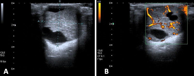
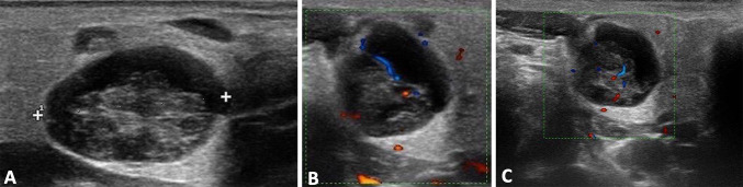
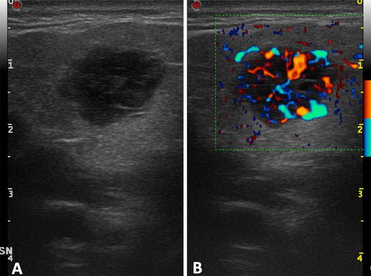
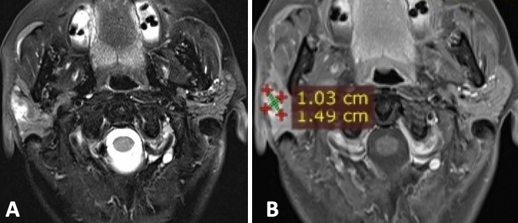
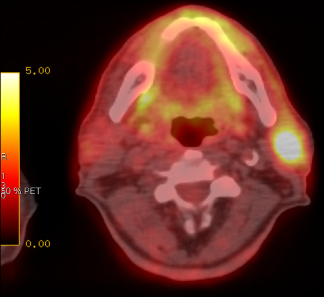
Parotid gland oncocytoma (PGO) is a rare benign epithelial tumor that usually occurs in the elderly population. The most common clinical presentation is a painless, slow-growing, non-tender, lobulated, and mobile mass. Histologically, it is composed of monotonous sheets of epithelial cells (oncocytes) with a central scar. The cross-sectional appearance is not specific, and it overlaps with other parotid lesions. On ultrasound (US), oncocytoma appears as an ovoid, well-defined, homogeneous, and hypoechoic lesion. Cystic and hemorrhagic areas as well as intralesional fat may be observed. Doppler analysis shows intratumoral vessels, sometimes with a spoke-wheel pattern. The peak systolic flow is high (up to 100 cm/sec). Furthermore, oncocytoma is avid of FDG on a PET scan, as well as a malignant tumor. Thus, a combined clinical, imaging, and pathologic assessment is essential to establish the most accurate diagnosis and plan the best treatment. US, combined with Doppler techniques, can play an important role in suggesting the diagnosis and confirming it through percutaneous sampling. The purpose of this review is to show the imaging findings in PGO, with special emphasis on the US appearance.
Keywords: Color doppler; Oncocytoma; Parotid gland; Tumor; Ultrasound.
© 2020. Società Italiana di Ultrasonologia in Medicina e Biologia (SIUMB).
Conflict of interest statement
We confirm that this work is original and has not been published elsewhere nor is it currently under consideration for publication elsewhere. Publication is approved by all authors and by the responsible authorities where the work was carried out. Each author have participated sufficiently in any submission to take public responsibility for its content. The authors have no conflicts of interest.
Figures

References
-
- Talas D, Go K. Incidental deep lobe parotid gland oncocytic neoplasms in an operated larynx cancer patient. Oral Oncol Extra. 2006;42(6):235–240. doi: 10.1016/j.ooe.2006.01.003. - DOI
Publication types
MeSH terms
LinkOut - more resources
Full Text Sources
