Review
. 2020 Nov;48(8):1043-1056.
doi: 10.1111/ceo.13834. Epub 2020 Aug 17.

Retinal pigment epithelium and age-related macular degeneration: A review of major disease mechanisms

Affiliations
Review

Retinal pigment epithelium and age-related macular degeneration: A review of major disease mechanisms

Shreya Somasundaran et al. Clin Exp Ophthalmol. 2020 Nov.

Abstract

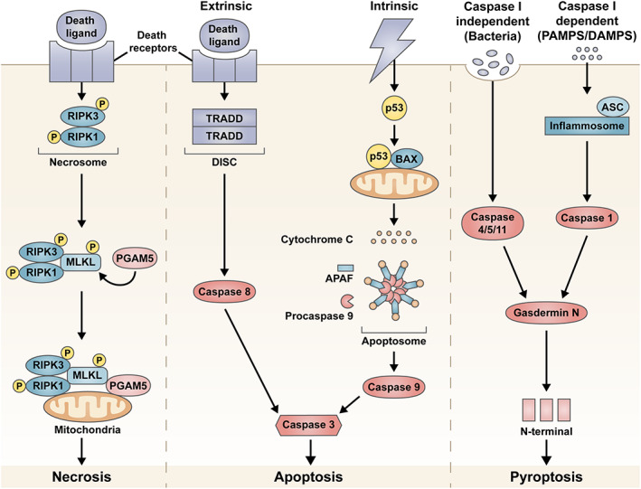
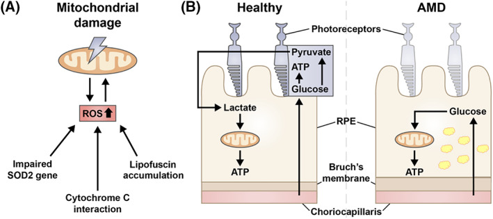
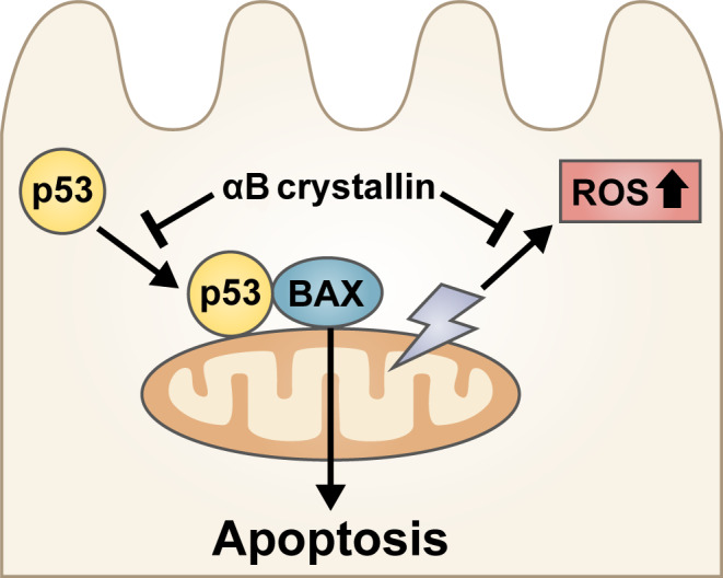
Age-related macular degeneration (AMD) is a progressive degenerative disease that is the leading cause of vision loss in the elderly population. Degeneration/dysregulation of the retinal pigment epithelium (RPE), a supportive monolayer of cells underlying the photoreceptors, is commonly seen in patients with AMD. While treatment exists for the neovascular/wet form of AMD, there is currently no cure for the non-exudative/dry form of AMD, making it imperative to understand the pathogenesis of this disease. Although our understanding of the aetiology of AMD has increased over the years, the underlying disease mechanism has not yet been identified, mainly due to the multifactorial nature of this disease. Herein, we review some of the commonly proposed degeneration pathways of RPE cells and their role in the pathogenesis of AMD; including activation of the complement cascade, oxidative stress-induced cell death mechanisms, dysfunctional mitochondria and the role of crystallins in AMD disease progression.

Keywords: age-related macular degeneration; apoptosis; eye; necrosis; retinal pigment epithelium.


Figures

FIGURE 1
FIGURE 1
Overview of the cell death pathways of necrosis, apoptosis and pyroptosis proposed as the retinal pigment epithelium (RPE) cell death mechanism in age‐related macular degeneration (AMD). In necrosis, the necrosome forms a complex with other proteins and attaches to the mitochondrial membrane initiating cell death. Programmed cell death can be triggered using either the intrinsic or extrinsic pathways, which eventually culminates in the activation of caspase 3 and cell death via apoptosis. Pyroptosis can be activated in either caspase 1 dependent or independent manner. Both pathways lead to the generation of a N‐terminal fragment that triggers cell death
FIGURE 2
FIGURE 2
Mitochondria dysfunction in the retinal pigment epithelium (RPE) is another proposed mechanism in age‐related macular degeneration (AMD). Mitochondrial DNA damage is linked to an increase in reactive oxidation species (ROS) production. Although the causal relationship between these events is unclear, an impaired superoxide dismutase 2 gene, cytochrome c interaction and lipofuscin accumulation can lead to increase in ROS production levels within the cell (A). Dysfunctional mitochondria due to metabolic coupling can also be a potential mechanism for AMD, where photoreceptor cell death ensues due starvation, as glucose meant to be used by the neural retina is consumed by the RPE (B). Note the thickening of the Bruch's membrane and accumulation of lipofuscin (yellow granules) within the RPE during AMD
FIGURE 3
FIGURE 3
αB crystallin may confer a protective role in the pathogenesis of age‐related macular degeneration (AMD). αB crystallin could potentially inhibit reactive oxygen species (ROS) overexpression and apoptosis driven by p53 translocation to the mitochondria

References

    1. Gehrs KM, Anderson DH, Johnson LV, Hageman GS. Age‐related macular degeneration—emerging pathogenetic and therapeutic concepts. Ann Med. 2006;38(7):450‐471. - PMC - PubMed
    1. Galloway CA, Dalvi S, Hung SSC, et al. Drusen in patient‐derived hiPSC‐RPE models of macular dystrophies. Proc Natl Acad Sci U S A. 2017;114(39):E8214‐E8223. - PMC - PubMed
    1. Gao H, Hollyfield JG. Aging of the human retina. Differential loss of neurons and retinal pigment epithelial cells. Invest Ophthalmol Vis Sci. 1992;33(1):1‐17. - PubMed
    1. Eldred GE, Katz ML. Fluorophores of the human retinal pigment epithelium: separation and spectral characterization. Exp Eye Res. 1988;47(1):71‐86. - PubMed
    1. Ugarte M, Hussain AA, Marshall J. An experimental study of the elastic properties of the human Bruch's membrane‐choroid complex: relevance to ageing. Br J Ophthalmol. 2006;90(5):621‐626. - PMC - PubMed