Review
. 2020 Sep:62:101128.
doi: 10.1016/j.arr.2020.101128. Epub 2020 Jul 23.

Inter and Intracellular mitochondrial trafficking in health and disease

Affiliations
Review

Inter and Intracellular mitochondrial trafficking in health and disease

Santhanam Shanmughapriya et al. Ageing Res Rev. 2020 Sep.

Abstract

Neurons and glia maintain central nervous system (CNS) homeostasis through diverse mechanisms of intra- and intercellular signaling. Some of these interactions include the exchange of soluble factors between cells via direct cell-to-cell contact for both short and long-distance transfer of biological materials. Transcellular transfer of mitochondria has emerged as a key example of this communication. This transcellular transfer of mitochondria are dynamically involved in the cellular and tissue response to CNS injury and play beneficial roles in recovery. This review highlights recent research addressing the cause and effect of intra- and intercellular mitochondrial transfer with a specific focus on the future of mitochondrial transplantation therapy. We believe that mitochondrial transfer plays a crucial role during bioenergetic crisis/deficit, but the quality, quantity and mode of mitochondrial transfer determines the protective capacity for the receiving cells. Mitochondrial transplantation is a new treatment paradigm and will overcome the major bottleneck of traditional approach of correcting mitochondria-related disorders.

Keywords: Extracellular vesicles; Kinesin; Miro; Mitochondrial extrusion; Mitochondrial transplantation; TRAK; Tunneling nanotubes.


Conflict of interest statement

Conflict of Interest

No potential conflict of interest was reported by the authors

Figures

Fig. 1.
Fig. 1.. Key components of the Motor-adaptor-receptor complex for intracellular mitochondrial movement.
Axonal microtubule (MT) are uniformly arranged with their plus end (+end) directed distally and minus end (−end) towards soma. (A) Plus-end directed anterograde transport (−end to +end) of mitochondria is mediated by the Kinesin motor. The N-terminal domain the motor domain with ATPase activity and binds directly the MT. The C-terminal is the cargo binding domain. Milton (or TRAK1/2) is the motor adaptor protein that links Miro (present in the outer mitochondrial membrane) to the cargo binding domain of Kinesin. (B) Minus-end directed retrograde transport (+end to −end) of mitochondria is mediated by the cytoplasmic MT-based dynein motor. Dynein contains multiple subunits including two catalytic heavy chains (DHC), several intermediate chains (DIC) and light chains (DLC). DHC is the motor domain required for dynein movement. Dynactin is a large protein complex that interacts with dynein and MT through the p150Glued subunit. Both dynein and dynactin together drive the retrograde mitochondrial movement. (C and D) Actin filament based short distance mitochondrial transport is mediated by myosin. Myosin is a two-headed motor protein with a unique globular tail domain that can interact directly with kinesin motor (C), or with DLC (D) raising the possibility of this motor to facilitate both long-range transport along MT and short-distance transport along actin filaments. (E) Myo19 was identified as the mitochondria associated myosin. Myo19 directly interacts with Miro through its C-terminal fragment of the tail region.
Fig. 2.
Fig. 2.. Molecular regulation of mitochondrial movement.
(A) Dissociation of motor/adaptor/receptor complex. Binding of Ca2+ to the Miro EF hands facilitate the dissociation of either Miro/TRAK or KIF5/TRAK interaction, thereby uncoupling the KIF5 from the mitochondrial surface. (B) Switching of kinesin from active to inactive state by directly binding to Miro. KIF5 switches from the active state to an inactive state by binding to the Ca2+ bound Miro through the head domain of kinesin, thus preventing it from binding to microtubules. (C) Irreversible removal of Miro from the complex. PINK1 binds to and phosphorylates Miro to signal Parkin. The consequence of activating the PINK/Parkin pathway is the proteasome-dependent degradation of Miro and consequent release of KIF5/TRAK complex from the mitochondrial surface. (D) Mitochondrial docking by syntaphilin. Syntaphilin (SPH) acts as a static anchor specific for axonal mitochondria. Depending on the energy requirement mitochondria binds the microtubules through the protein syntaphilin and docks at the site of high energy demand.
Fig. 3.
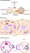
Fig. 3.. Mode of intercellular mitochondrial transfer.
(A) Tunneling nanotubes.P53-mediated activation of caspase 3 cleaves S100A4, creating a gradient of low levels of S100A4 in initiating or recipient cells (injured cells) towards a higher concentration in target or donor cells (healthy cells). It is suggested that RAGE in recipient cells act as the putative receptor for the high concentration of S100A4 in donor cells and this co-ordination of S100A4 and RAGE guides TNT direction. Mitochondria transfer through nanotunnels is believed to be facilitated by Actin/Miro-based transport machinery. (B) Gap junction. Connexin, specifically connexin 43 oligomerizes to form a channel at the gap junction which facilitates the transfer of mitochondria. Mechanistically, it was shown that ROS-induced oxidative stress regulates the opening of connexin channels in a system mediated by phosphoinositide 3-kinase (PI3K). (C) Extracellular vesicle. The multivesicular bodies (MVB) release extracellular vesicles (EVs) from the donor cell. These released EVs can fuse with recipient cells (1) or engulfed (2) to release the EVs’ content

References

    1. Abounit S, Zurzolo C, 2012. Wiring through tunneling nanotubes--from electrical signals to organelle transfer. J Cell Sci 125, 1089–1098. - PubMed
    1. Ahmad T, Mukherjee S, Pattnaik B, Kumar M, Singh S, Kumar M, Rehman R, Tiwari BK, Jha KA, Barhanpurkar AP, Wani MR, Roy SS, Mabalirajan U, Ghosh B, Agrawal A, 2014. Miro1 regulates intercellular mitochondrial transport & enhances mesenchymal stem cell rescue efficacy. EMBO J 33, 994–1010. - PMC - PubMed
    1. Al Amir Dache Z, Otandault A, Tanos R, Pastor B, Meddeb R, Sanchez C, Arena G, Lasorsa L, Bennett A, Grange T, El Messaoudi S, Mazard T, Prevostel C, Thierry AR, 2020. Blood contains circulating cell-free respiratory competent mitochondria. FASEB J 34, 3616–3630. - PubMed
    1. Araki T, 1977. Freezing injury in mitochondrial membranes. II. Degradation of phospholipid in rabbit liver mitochondria during freezing and storage at low temperatures. Cryobiology 14, 151–159. - PubMed
    1. Aspenstrom P, Ruusala A, Pacholsky D, 2007. Taking Rho GTPases to the next level: the cellular functions of atypical Rho GTPases. Exp Cell Res 313, 3673–3679. - PubMed

Publication types