. 2020 Oct:158:149-161.
doi: 10.1016/j.freeradbiomed.2020.07.006. Epub 2020 Jul 23.

Membrane insertion exacerbates the α-Synuclein-Cu(II) dopamine oxidase activity: Metallothionein-3 targets and silences all α-synuclein-Cu(II) complexes

Affiliations

Membrane insertion exacerbates the α-Synuclein-Cu(II) dopamine oxidase activity: Metallothionein-3 targets and silences all α-synuclein-Cu(II) complexes

Jenifer S Calvo et al. Free Radic Biol Med. 2020 Oct.

Abstract

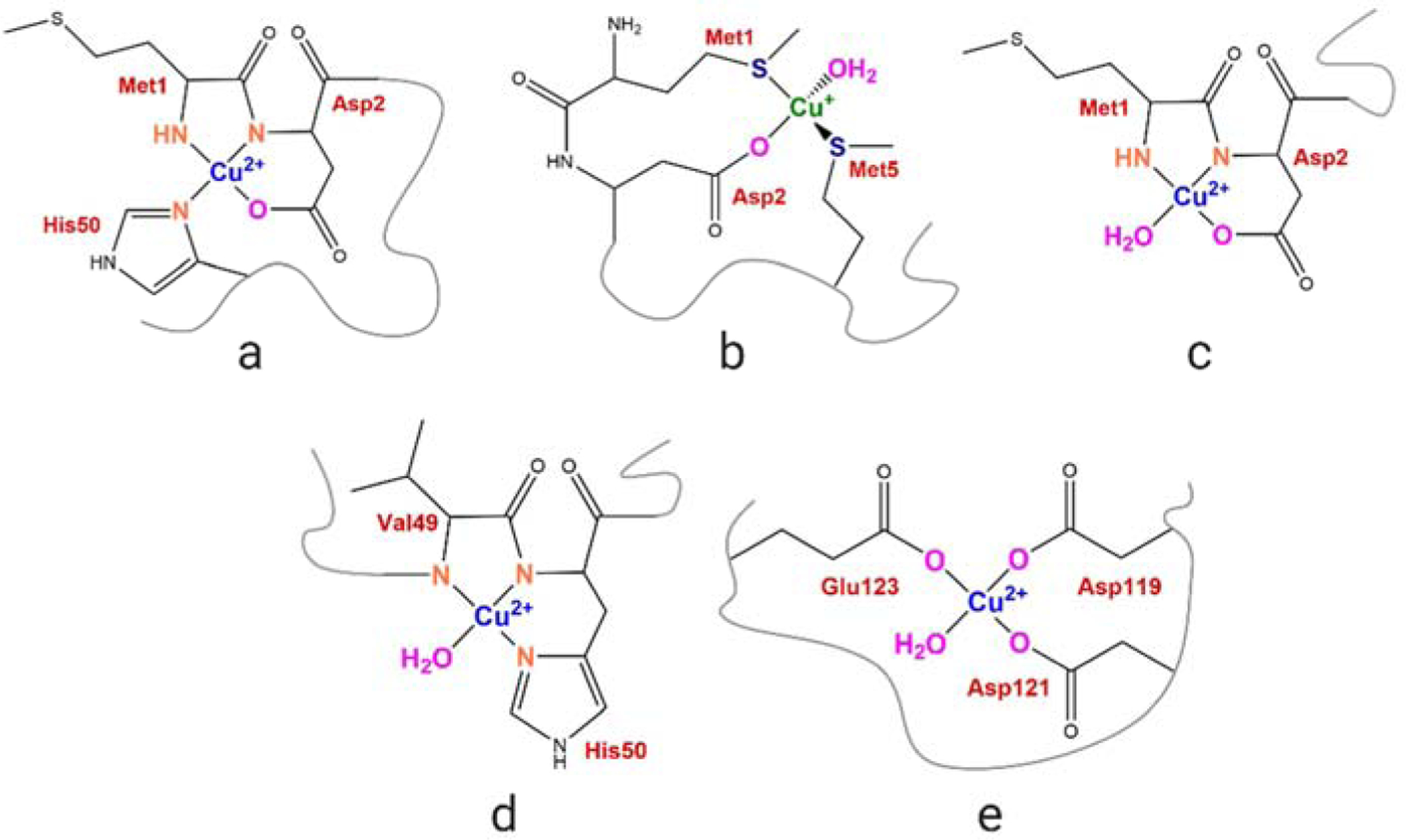
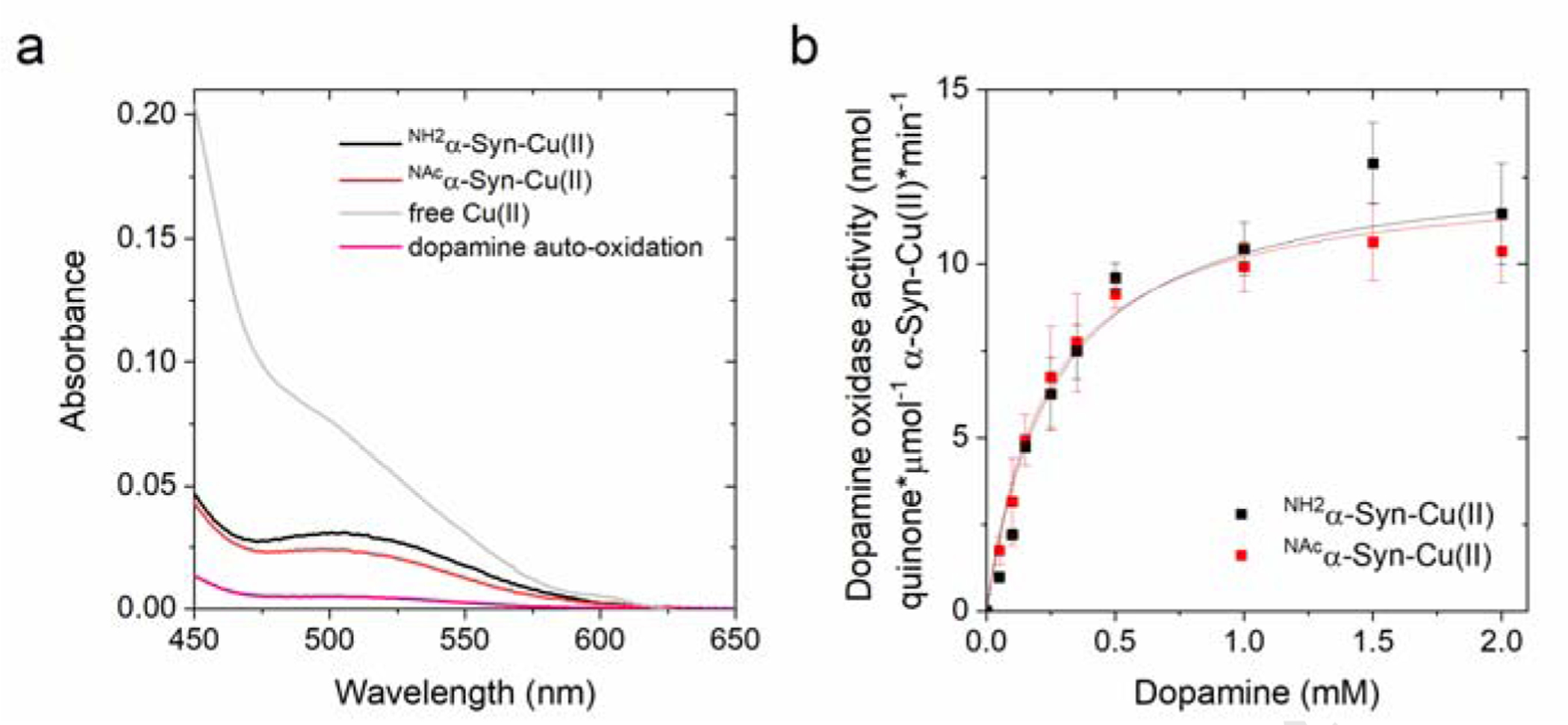
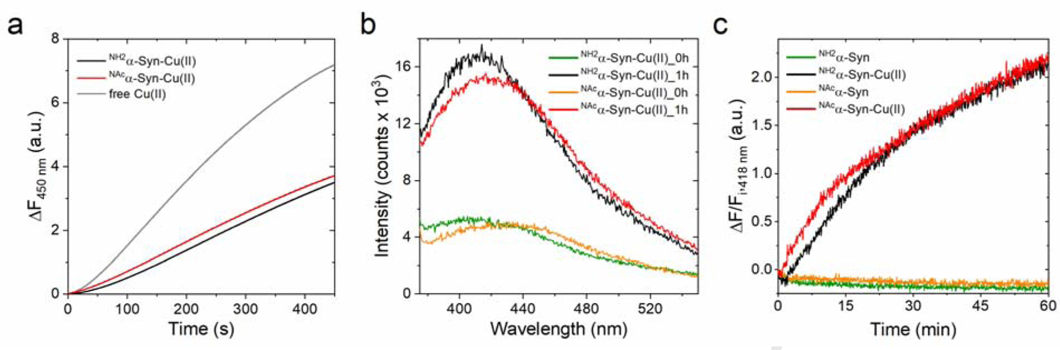
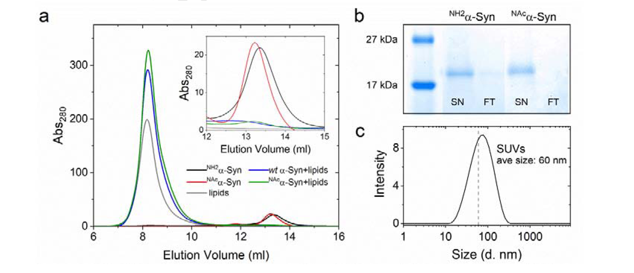
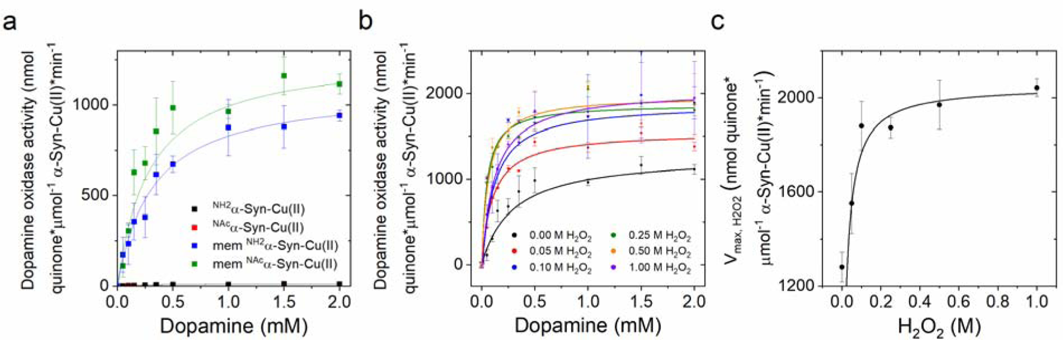
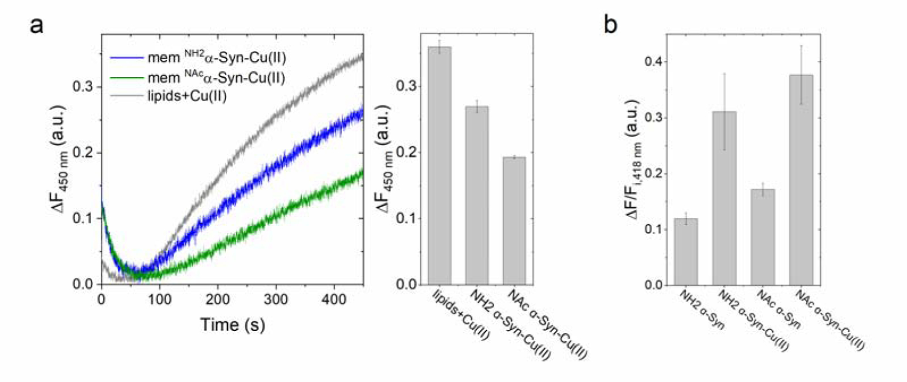
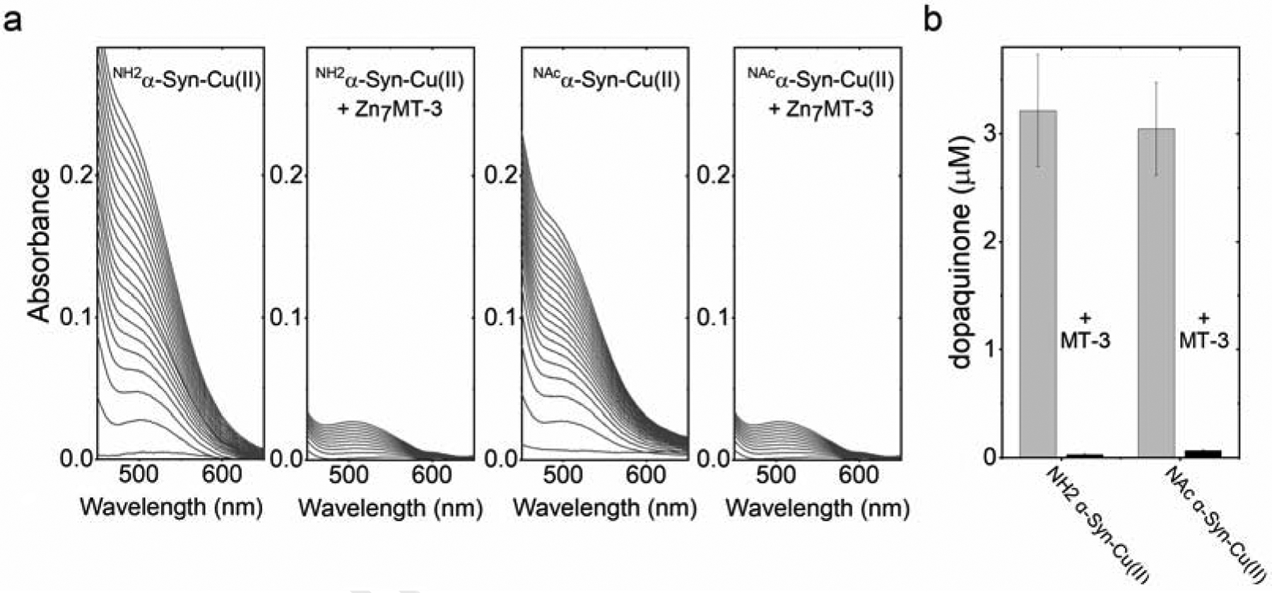
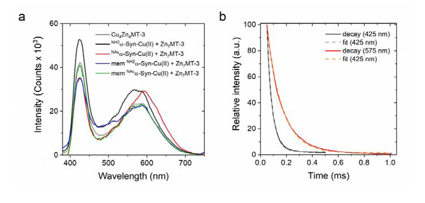
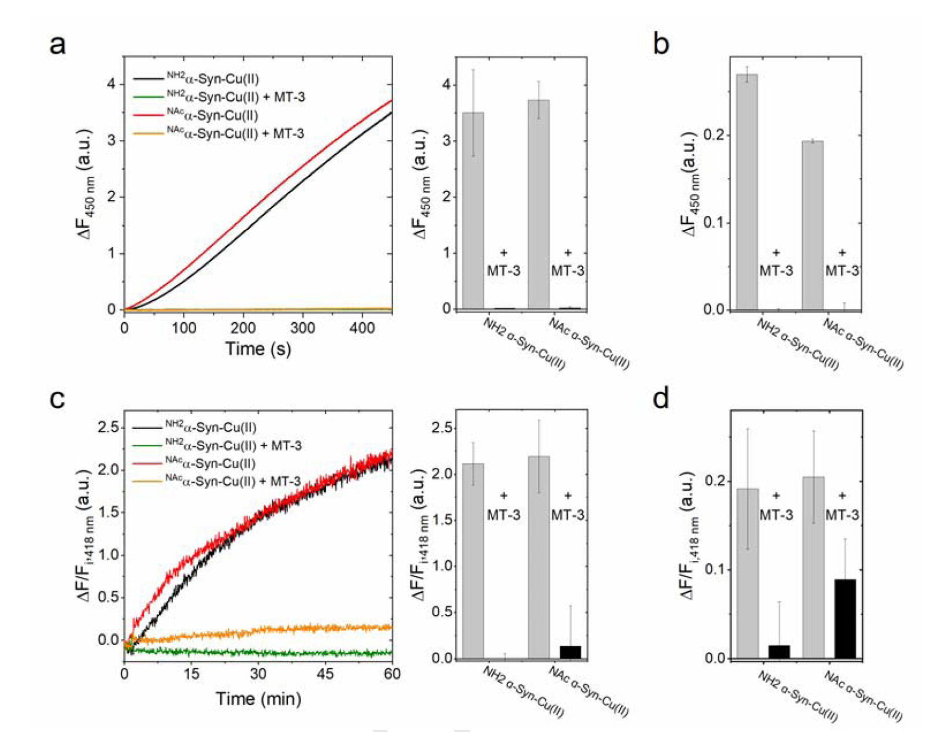
Copper binding to α-synuclein (α-Syn), the major component of intracellular Lewy body inclusions in substantia nigra dopaminergic neurons, potentiate its toxic redox-reactivity and plays a detrimental role in the etiology of Parkinson disease (PD). Soluble α-synuclein-Cu(II) complexes possess dopamine oxidase activity and catalyze ROS production in the presence of biological reducing agents via Cu(II)/Cu(I) redox cycling. These metal-centered redox reactivities harmfully promote the oxidation and oligomerization of α-Syn. While this chemistry has been investigated on recombinantly expressed soluble α-Syn, in vivo, α-Syn is acetylated at its N-terminus and is present in equilibrium between soluble and membrane-bound forms. This post-translational modification and membrane-binding alter the Cu(II) coordination environment and binding modes and are expected to affect the α-Syn-Cu(II) reactivity. In this work, we first investigated the reactivity of acetylated and membrane-bound complexes, and subsequently addressed whether the brain metalloprotein Zn7-metallothionein-3 (Zn7MT-3) possesses a multifaceted-role in targeting these aberrant copper interactions and consequent reactivity. Through biochemical characterization of the reactivity of the non-acetylated/N-terminally acetylated soluble or membrane-bound α-Syn-Cu(II) complexes towards dopamine, oxygen, and ascorbate, we reveal that membrane insertion dramatically exacerbates the catechol oxidase-like reactivity of α-Syn-Cu(II) as a result of a change in the Cu(II) coordination environment, thereby potentiating its toxicity. Moreover, we show that Zn7MT-3 can efficiently target all α-Syn-Cu(II) complexes through Cu(II) removal, preventing their deleterious redox activities. We demonstrate that the Cu(II) reduction by the thiolate ligands of Zn7MT-3 and the formation of Cu(I)4Zn4MT-3 featuring an unusual redox-inert Cu(I)4-thiolate cluster is the molecular mechanism responsible for the protective effect exerted by MT-3 towards α-Syn-Cu(II). This work provides the molecular basis for new therapeutic interventions to control the deleterious bioinorganic chemistry of α-Syn-Cu(II).

Keywords: Alpha-synuclein; Copper dysregulation; Dopamine oxidation; Metallothionein-3; Parkinson's disease; Reactive oxygen species.


Figures

Figure 1.
Figure 1.
Proposed copper coordination modes in α-Syn. (a) Cu(II) coordination at the high-affinity N-terminal site in soluble α-Syn at physiological pH; (b) Cu(I) coordination at the N-terminal site; (c) high affinity N-terminal site in membrane-bound form; (d) NAcα-Syn high-affinity site centered at His50; and (E) low-affinity C-terminal site.
Figure 2.
Figure 2.
(a) Absorption spectra recorded after the reaction of NH2α-Syn-Cu(II) (10 μM; black), NAcα-Syn-Cu(II) (10 μM; red), or free Cu(II) (10 μM; gray) with dopamine (1 mM) in 20 mM N-ethylmorpholine/100 mM NaCl, pH 7.4 at 25°C, in the presence of MBTH (2 mM). Auto-oxidation of dopamine (1 mM) is plotted in pink. (b) Concentration-dependent dopamine oxidase activity of NH2α-Syn-Cu(II) (10 μM; black) or NAcα-Syn-Cu(II) (10 μM; red) in 20 mM N-ethylmorpholine/100 mM NaCl, pH 7.4, determined using MBTH (2 mM) to quantify the dopamine ortho-quinone formed after 120 s reaction (25°C).
Figure 3.
Figure 3.
(a) Ascorbate-driven hydroxyl radical production by Cu(II) (5 μM; gray), NH2α-Syn-Cu(II) (5 μM; black), or NAcα-Syn-Cu(II) (5 μM; red) in the presence of ascorbate (600 μM) and 3-CCA (400 μM), determined by monitoring the formation of the fluorescent product 7-OH-CCA for 450 s at 37°C (λex=395 nm; λem=450 nm). (b) Dityrosine emission spectra (λex=325 nm) recorded forNH2α-Syn-Cu(II) (10 μM; green and black) and NAcα-Syn (10 μM; orange and red) before and after 1-h reaction with 1 mM ascorbate (37°C). (c) Kinetic traces monitoring dityrosine formation at 418 nm (λex=325 nm) in NH2α-Syn (green), NH2α-Syn-Cu(II) (black), NAcα-Syn (orange), and NAcα-Syn-Cu(II) (red). Dityrosine formation is reported as the difference between final and initial fluorescence at 418 nm (ΔF) over the initial fluorescence (Fi).
Figure 4.
Figure 4.
(a) Size exclusion chromatogram monitoring membrane insertion of soluble NH2α-Syn (15 μM; black, blue after insertion) or NAcα-Syn (red, green after insertion) using a Superdex 200 column. Lipids (gray) elute at the void volume (~8.2 ml) while soluble α-Syn elutes at 13.3 ml (black and red). Inset: Enlarged portion of the chromatogram showing soluble α-Syn elution. (b) SDS-PAGE of membrane-bound NH2α-Syn and NAcα-Syn, generated after removing soluble protein using 50-kDa MWCO filters (SN: supernatant; FT: filtrate). (c) Dynamic light scattering size distribution analysis of lipid vesicles (0.1 mg/ml lipids) generated by sonication, indicating the formation of SUVs.
Figure 5.
Figure 5.
(a) Concentration-dependent dopamine oxidase activity of soluble or membrane-bound NH2α-Syn-Cu(II) (10 μM; black and blue, respectively) and NAcα-Syn-Cu(II) (10 μM; red and green, respectively) in 20 mM N-ethylmorpholine/100 mM NaCl, pH 7.4, determined using MBTH (2mM) to quantify the dopaquinone formed. Values were obtained after 120 s reaction for soluble forms and 20 s for the membrane-bound forms (25°C). (b) Concentration-dependent dopamine oxidase activity of membrane-bound NAcα-Syn-Cu(II) (10 μM) in 20 mM N-ethylmorpholine/100 mM NaCl, pH 7.4 at increasing H2O2 concentrations (0 – 1 M), determined using 2 mM MBTH to quantify the dopaquinone formation (20 s, 25°C). (c) Michaelis-Menten analysis to determine Vmax, H2O2 as a function of H2O2 concentrations.
Figure 6.
Figure 6.
(a) Ascorbate-driven hydroxyl radical production of membrane-bound NH2α-Syn-Cu(II) (5 μM; blue), membrane-bound NAcα-Syn-Cu(II) (5 μM; green), or Cu(II) bound to lipids (5 μM; gray), determined in the presence of 600 μM ascorbate and 400 μM 3-CCA by monitoring the formation of fluorescent product 7-OH-CCA (λex=395 nm; λem=450 nm; 450 s; 37°C.) (b) Ascorbate-driven dityrosine formation of membrane-bound NH2α-Syn-Cu(II) or NAcα-Syn-Cu(II) (5 μM), determined by monitoring dityrosine fluorescence at 418 nm (λex=325 nm) upon incubation with 3 mM ascorbate for 3 h (37°C). Dityrosine formation is reported as the difference between final and initial fluorescence at 418 nm (ΔF) divided by initial fluorescence (Fi).
Figure 7.
Figure 7.
(a) Catalytic dopamine (1 mM) oxidation by NH2α-Syn-Cu(II) or NAcα-Syn-Cu(II) (10 μM) in 20 mM N-ethylmorpholine/100 mM NaCl, pH 7.4, in the presence 2 mM MBTH (100 min, 25°C). The dopamine oxidase activity is quenched upon addition of Zn7MT-3 (0.25 eq., 1 h) prior to the addition of dopamine. (b) Quenching of the dopamine oxidase activity of membrane-bound NH2α-Syn-Cu(II) or NAcα-Syn-Cu(II) (10 μM) upon reaction with Zn7MT-3 (0.25 eq.). The dopaquinone formed was quantified after 20 s (25°C) using 2 mM MBTH.
Figure 8.
Figure 8.
(a) Low-temperature (77 K) luminescence emission spectra of the products of the reaction (1 h; 25°C) between soluble and membrane-bound NH2α-Syn-Cu(II) (10 μM; black and blue, respectively) or NAcα-Syn-Cu(II) (10 μM; red and green, respectively) with Zn7MT-3 (2.5 μM). The reference spectra of Cu(I)4Zn(II)4MT-3 (2.5 μM) is plotted for comparison (gray). (b) Emission lifetime determination of the product of reaction between membrane-bound NAcα-Syn-Cu(II) and Zn7MT-3 at 425 nm (black) and 575 nm (red). Lifetime values were determined by fitting using single exponential decay function (gray and orange, respectively).
Figure 9.
Figure 9.
(a) Quenching of ascorbate-driven hydroxyl radical production of 5 μM soluble NH2α-Syn-Cu(II) (black) and NAcα-Syn-Cu(II) (red) by 0.25 eq. Zn7MT-3 (green and orange lines, respectively) incubated for 1 h before reaction with 1 mM ascorbate. Hydroxyl radical production was monitored by following the formation of the fluorescent product 7-OH-CCA (λex=395 nm; λem=450 nm) for 450 s (37°C). (b) Hydroxyl radical quenching by Zn7MT-3 (1.25 μM) in the membrane-bound α-Syn-Cu(II) forms (5 μM). (c) Quenching of ascorbate-driven dityrosine formation of soluble NH2α-Syn-Cu(II) (black) and NAcα-Syn-Cu(II) (red) by Zn7MT-3 (green and orange lines, respectively), incubated for 1 h before reaction was initiated by addition of ascorbate (1 mM). Dityrosine formation was determined by monitoring dityrosine fluorescence at 418 nm (λex=325 nm) after 1 h reaction (37°C), reported as the difference between final and initial fluorescence (ΔF) divided by initial fluorescence (Fi). (d) Quenching of dityrosine formation by Zn7MT-3 (1.25 μM) in the membrane-bound α-Syn-Cu(II) forms (5 μM), after following the reaction for 3 h upon the addition of ascorbate (3 mM). Values are corrected for ΔF/Fi, 418nm of membrane-bound α-Syn forms in the absence Cu(II).

References

    1. de Lau LML and Breteler MMB, Epidemiology of Parkinson’s disease, Lancet Neurol, 5, 6, 525–535, 2006, doi: 10.1016/S1474-4422(06)70471-9. - DOI - PubMed
    1. Spillantini MG, Schmidt ML, Lee VMY, Trojanowski JQ, Jakes R, and Goedert M, α-Synuclein in Lewy bodies, Nature, 388, 6645, 839–840, 1997, doi: 10.1038/42166. - DOI - PubMed
    1. Spillantini MG, Crowther RA, Jakes R, Hasegawa M, and Goedert M, α-Synuclein in filamentous inclusions of Lewy bodies from Parkinson’s disease and dementia with Lewy bodies, Proc. Natl. Acad. Sci, 95, 11, 6469–6473, 1998, doi: 10.1073/pnas.95.11.6469. - DOI - PMC - PubMed
    1. Stefanis L, α-Synuclein in Parkinson’s disease, Cold Spring Harb. Perspect. Med, 2, 2, 1–23, 2012, doi: 10.1101/cshperspect.a009399. - DOI - PMC - PubMed
    1. Anderson JP et al., Phosphorylation of Ser-129 is the dominant pathological modification of α-synuclein in familial and sporadic lewy body disease, J. Biol. Chem, 281, 40, 29739–29752, 2006, doi: 10.1074/jbc.M600933200. - DOI - PubMed

Publication types