

The Histological Basis of Frank's Sign
- PMID: 32712879
- PMCID: PMC8134579
- DOI: 10.1007/s12105-020-01205-4
The Histological Basis of Frank's Sign
Abstract
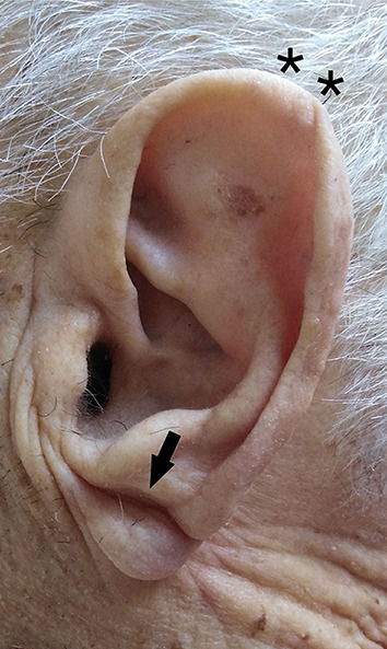
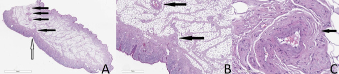
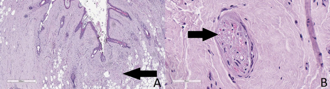
Frank's sign is a diagonal crease of the ear lobe, supposedly related to cardiac pathology, and has strongly been associated with coronary artery atherosclerosis. A total of 45 consecutive adult patients referred for autopsy in a one-and-a-half-year period were extensively studied. Samples from both the ear lobes were obtained for histopathology, as well as cardiac samples from all four cardiac compartments. When compared patients with Frank's sign and those without it had no statistical difference in age (p = 0.0575). There was however a statistically significant increased cardiac weight (p = 0.0005), left ventricular wall thickness (p = 0.0002), and right ventricular wall thickness (p = 0.0043). Histopathology obtained from the ear lobes revealed myoelastofibrosis in an arterial vessel, located at the base of the crease, diffuse fibrosis, and Wallerian-like degeneration, with eosinophilic inclusions in the peripheral nerves. These changes suggest a time-related progression of the crease-associated changes. Our data suggest a significant correlation between the morphological changes of the myocardium and the presence of the ear lobe creases, with arterial myoelastofibrosis, Wallerian-like degeneration in peripheral nerves and deep tissue fibrosis found in the base of the crease.
Keywords: Fibrosis; Frank’s sign; Heart failure; Hypoxia; Neural degeneration; Pathology.
Conflict of interest statement
The authors declare that they have no competing interest.
Figures
References
MeSH terms
LinkOut - more resources
Full Text Sources
Medical
