Review
. 2021 Jun;15(2):657-662.
doi: 10.1007/s12105-020-01206-3. Epub 2020 Jul 25.

Kimura Disease Presenting with Buccal Mass: A Case Report and Literature Review

Affiliations
Review

Kimura Disease Presenting with Buccal Mass: A Case Report and Literature Review

Ryosuke Sato et al. Head Neck Pathol. 2021 Jun.

Abstract

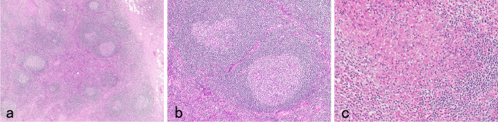
A 52-year-old man with a 2-year history of left buccal swelling was admitted to our department. An elastic hard oral mass was palpated under the intact buccal mucosa. A CT scan with enhancement revealed a solid mass measuring 2.0 × 1.5 × 1.3 cm between the left masseter muscle and the maxilla. Laboratory examination showed elevated peripheral blood eosinophil count of 1070/μL (12.3%) and serum immunoglobulin (Ig)E level of 1374 IU/mL. Histologic examination of transorally excised mass revealed lymphoid follicular hyperplasia with reactive germinal centers and eosinophilic infiltration with eosinophilic micro-abscesses in the germinal centers. Abundant IgE deposition in a reticular fashion was observed in the germinal centers and c-kit positive mast cells was observed in the paracortical area in the excised mass. The patient was diagnosed with Kimura disease (KD) and treated with oral prednisolone, tapering from 10 mg/day for approximately 8 months. Eosinophil count and serum IgE level decreased to 435/μL (5%) and 520 IU/dL, respectively. He is free from symptoms at the time of this submission. KD, a rare, benign, and chronic inflammatory disorder, occurs predominantly in young male adults in Asia. Patients with KD who presents with buccal mass are relatively rare. Immunohistologic analyses suggested that an allergic reaction played an important role in the etiology of KD in this case.

Keywords: Buccal mass; Eosinophil; Immunoglobulin E; Kimura disease; Steroid.


Conflict of interest statement

The authors declare that there are no actual or potential conflicts of interest in relation to this article.

Figures

Fig. 1
Fig. 1
Swelling of the left buccal area was observed (a). An elastic hard oral mass was palpated under the intact buccal mucosa (b)
Fig. 2
Fig. 2
CT scan with enhancement revealed a solid mass measuring 2 × 1.5 × 1.3 cm between the left masseter muscle and the maxilla (triangle), with edema around the mass (a). MRI revealed a solid mass in the left buccal space, which exhibited a hypo-intense signal on T1-weighted image (b) and iso-intense signal on T2-weighted image (c)
Fig. 3
Fig. 3
Resolution of swelling around the left buccal area was observed (a) and the mass could not be detected on T2-weighted MRI (b) at 8 months after the start of prednisolone treatment
Fig. 4
Fig. 4
Histologic features with hematoxylin eosin staining of a specimen excised from the buccal mass. Lymphoid follicular hyperplasia with reactive germinal centers, an increase in vessels and inflammatory infiltrate within and around the lymphoid follicles (a, × 20, b, × 100) and intense eosinophilic infiltration with eosinophilic micro-abscesses in the degenerated germinal centers were observed (c, × 200)
Fig. 5
Fig. 5
Immunohistologic features of a specimen excised from the buccal mass. CD20-positive B cells were present in all areas, including the lymphoid follicles and paracortical area (a). CD3-positive T cells were present in the germinal centers and paracortical area but scarce in the mantle zones (b). Abundant IgE deposition in a reticular fashion was observed in the germinal centers (c). Only a few IgG4-positive cells were observed in the germinal centers (d). C-kit–positive cells were scattered in the paracortical area (e). The c-kit–positive cells, which appeared round or polyhedral in shape and were larger in size compared to other lymphocytes, were regarded as mast cells (f). Magnification: × 100 (ae), × 400 (f)

References

    1. Chen H, Thompson LD, Aguilera NS, Abbondanzo SL. Kimura disease: a clinicopathologic study of 21 cases. Am J Surg Pathol. 2004;28(4):505–513. doi: 10.1097/00000478-200404000-00010. - DOI - PubMed
    1. Kimura T, Yoshimura S, Ishikawa H. Unusual granuration combained with hyperplastic change of lymphoid tissues. Trans Soc Pathol Jpn. 1948;37:179–180.
    1. Ishii M. Kimura disease (in Japanease) ORL Tokyo. 1982;25(4):407–416.
    1. Iida S, Fukuda Y, Ueda T, et al. Kimura's disease: report of a case with presentation in the cheek and upper eyelid. J Oral Maxillofac Surg. 2005;63(5):690–693. doi: 10.1016/j.joms.2005.01.011. - DOI - PubMed
    1. Lee SJ, Song JH, Kim SD. Kimura's disease involving the ipsilateral face and extraocular muscles. Korean J Ophthalmol. 2009;23(3):219–223. doi: 10.3341/kjo.2009.23.3.219. - DOI - PMC - PubMed

LinkOut - more resources