Review
. 2020 Sep 14;375(1807):20190387.
doi: 10.1098/rstb.2019.0387. Epub 2020 Jul 27.

Rules of collective migration: from the wildebeest to the neural crest

Affiliations
Review

Rules of collective migration: from the wildebeest to the neural crest

Adam Shellard et al. Philos Trans R Soc Lond B Biol Sci. .

Abstract

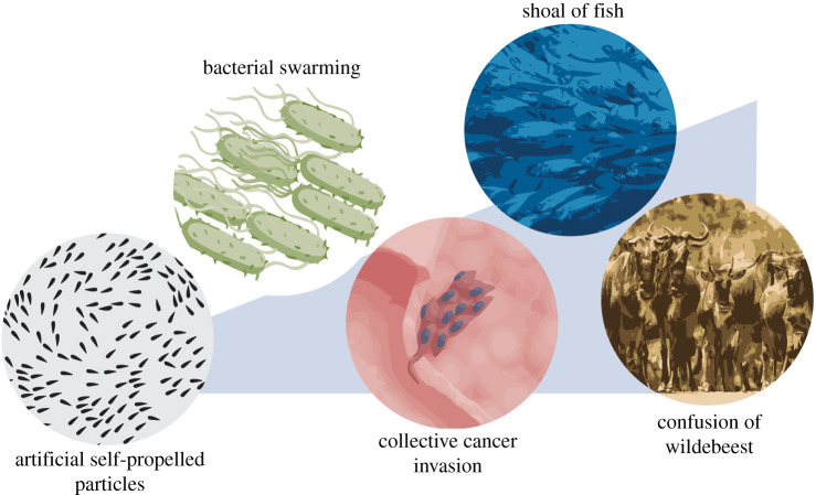
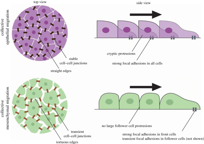
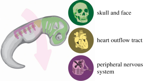
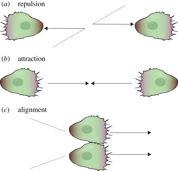
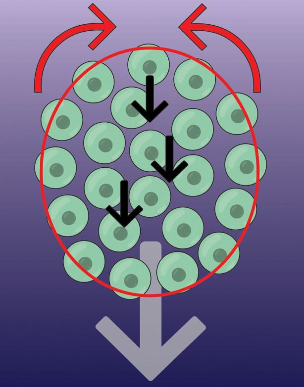
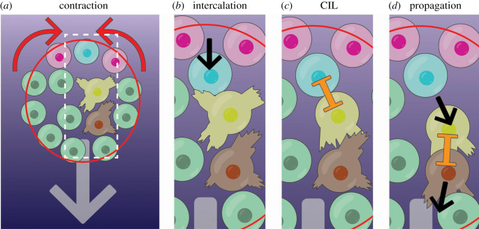
Collective migration, the movement of groups in which individuals affect the behaviour of one another, occurs at practically every scale, from bacteria up to whole species' populations. Universal principles of collective movement can be applied at all levels. In this review, we will describe the rules governing collective motility, with a specific focus on the neural crest, an embryonic stem cell population that undergoes extensive collective migration during development. We will discuss how the underlying principles of individual cell behaviour, and those that emerge from a supracellular scale, can explain collective migration. This article is part of the theme issue 'Multi-scale analysis and modelling of collective migration in biological systems'.

Keywords: alignment; co-attraction; collective migration; contact inhibition of locomotion; neural crest; supracellular.


Conflict of interest statement

We declare we have no competing interests.

Figures

Figure 1.
Figure 1.
Collective migration at all scales. Collective migration is found at practically all levels, from self-propelled particles to bacteria, cancer and animals.
Figure 2.
Figure 2.
Collective migration of epithelial cells and mesenchymal cells. Epithelial migration can arise from an unjamming transition of quiescent epithelial sheets. Such unjammed, motile epithelia display packs of collectively migrating cells (purple cells), while maintaining strong intercellular junctions (dark brown rectangles) and epithelial markers, such as E-cadherin. Epithelial migration is also evident in wound healing. Leader cells tend to form large forward-facing protrusions, with follower cells also contributing significant traction forces to move the sheet forward (teal rectangles). Mesenchymal migration can arise from an epithelial-to-mesenchymal transition, in which cells (green cells) lose apicobasal polarity in favour of front-rear polarity, and intercellular adhesions become weaker and more transient (light brown rectangles), which is associated with a change in gene expression, such as E-cadherin being replaced by N-cadherin. Whereas leader cells form strong focal adhesions, follower cells do not. The black arrow indicates the direction of migration.
Figure 3.
Figure 3.
Neural crest migration. The neural crest form at the border of the neural plate (top of the embryo), and then collectively migrate (pink arrow) toward the pharyngeal arches (bottom of the embryo). The green areas correspond to cranial neural crest migration, the yellow to cardiac neural crest migration, and the purple to trunk neural crest migration, which must move over the somites (brown ovals). When they reach the pharyngeal arches, they differentiate into a variety of cell types and contribute to many tissues and organs, including the craniofacial structures, the outflow tract, dorsal root ganglia and the enteric nervous system.
Figure 4.
Figure 4.
Cellular mechanisms of the three ‘rules’ of collective migration. (a) Two colliding cells (black dotted lines) repolarize and move away from each other (black arrows) by contact inhibition of locomotion. Purple regions represent the local activity of Rac1 at the leading edge and brown regions represent the local activity of RhoA at the cell rear. (b) Two cells are mutually attracted by C3a-dependent chemotaxis. (c) Two colliding cells move together for a short period of time after colliding, and before repulsion. This is one mechanism that drives the alignment of cell motion.
Figure 5.
Figure 5.
Supracellular migration of the neural crest. A group of neural crest cells (green circles) migrate forward by chemotaxis (grey arrow; chemotactic gradient is purple background). The mechanism for this directed movement relies on a supracellular contraction force at the rear (red arrows), driven by a pluricellular actomyosin cable (red). Contraction is inhibited by chemoattractant at the front. This rear force causes cells to intercalate forward (black arrows), moving to the front before becoming mechanically connected at the edge.
Figure 6.
Figure 6.
Stages of supracellular neural crest cell migration. (a) Cells at the edge of the cluster are linked by an actomyosin cable (red). The cable contracts at the rear (red cable, red arrows indicate contraction) but not at the front. (b) All cells at the rear are brought closer together, causing cells to intercalate forwards (blue cells intercalation as pink cells move closer together; movement is black arrow). (c) The intercalating cell (blue) makes contact with the unpolarized cell in front (yellow), causing it to polarize, producing protrusions forward. This occurs by CIL (orange inhibition symbol). (d) This cell (yellow) then moves forward, propagating the signal to the cells in front (brown cell), and so forth. Thus, an anterograde wave of aligned forward cell flow emanates from the rear of the cell cluster.

References

    1. Berdahl AM, Kao AB, Flack A, Westley PAH, Codling EA, Couzin ID, Dell AI, Biro D. 2018. Collective animal navigation and migratory culture: from theoretical models to empirical evidence. Phil. Trans. R. Soc. B 373, 20170009 (10.1098/rstb.2017.0009) - DOI - PMC - PubMed
    1. Chen C, Liu S, Shi XQ, Chate H, Wu YL. 2017. Weak synchronization and large-scale collective oscillation in dense bacterial suspensions. Nature 542, 210–214. (10.1038/nature20817) - DOI - PubMed
    1. Kearns DB. 2010. A field guide to bacterial swarming motility. Nat. Rev. Microbiol. 8, 634–644. (10.1038/nrmicro2405) - DOI - PMC - PubMed
    1. Ariel G, Rabani A, Benisty S, Partridge JD, Harshey RM, Be'er A. 2015. Swarming bacteria migrate by Levy Walk. Nat. Commun. 6, 8396 (10.1038/ncomms9396) - DOI - PMC - PubMed
    1. Friedl P, Locker J, Sahai E, Segall JE. 2012. Classifying collective cancer cell invasion. Nat. Cell Biol. 14, 777–783. (10.1038/ncb2548) - DOI - PubMed

Publication types

LinkOut - more resources