Collective dynamics of sperm cells
- PMID: 32713305
- PMCID: PMC7423380
- DOI: 10.1098/rstb.2019.0384
Collective dynamics of sperm cells
Abstract
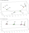
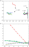
While only a single sperm may fertilize the egg, getting to the egg can be facilitated, and possibly enhanced, by sperm group dynamics. Examples range from the trains formed by wood mouse sperm to the bundles exhibited by echidna sperm. In addition, observations of wave-like patterns exhibited by ram semen are used to score prospective sample fertility for artificial insemination in agriculture. In this review, we discuss these experimental observations of collective dynamics, as well as describe recent mechanistic models that link the motion of individual sperm cells and their flagella to observed collective dynamics. Establishing this link in models involves negotiating the disparate time- and length scales involved, typically separated by a factor of 1000, to capture the dynamics at the greatest length scales affected by mechanisms at the shortest time scales. Finally, we provide some outlook on the subject, in particular, the open questions regarding how collective dynamics impacts fertility. This article is part of the theme issue 'Multi-scale analysis and modelling of collective migration in biological systems'.
Keywords: biological fluid dynamics; collective dynamics; sperm motility.
Conflict of interest statement
We declare we have no competing interests.
Figures


Similar articles
-
Relationship between in vitro sperm functional tests and in vivo fertility of rams following cervical artificial insemination of ewes with frozen-thawed semen.Theriogenology. 2008 Mar 1;69(4):513-22. doi: 10.1016/j.theriogenology.2007.12.003. Epub 2008 Jan 30. Theriogenology. 2008. PMID: 18248736
-
Post-thaw survival of ram spermatozoa and fertility after insemination as affected by prefreezing sperm concentration and extender composition.Theriogenology. 2001 Mar 15;55(5):1159-70. doi: 10.1016/s0093-691x(01)00474-5. Theriogenology. 2001. PMID: 11322242
-
Fertility of undiluted ram epididymal spermatozoa stored for several days at 4°C.Animal. 2015 Feb;9(2):313-9. doi: 10.1017/S1751731114002109. Epub 2014 Sep 25. Animal. 2015. PMID: 25252882
-
Improvement strategies in ovine artificial insemination.Reprod Domest Anim. 2006 Oct;41 Suppl 2:30-42. doi: 10.1111/j.1439-0531.2006.00767.x. Reprod Domest Anim. 2006. PMID: 16984467 Review.
-
Oviducal storage of spermatozoa in the turkey: its relevance to artificial insemination technology.Br Poult Sci. 1989 Jun;30(2):423-9. doi: 10.1080/00071668908417166. Br Poult Sci. 1989. PMID: 2670069 Review.
Cited by
-
Conservation Biology and Reproduction in a Time of Developmental Plasticity.Biomolecules. 2022 Sep 14;12(9):1297. doi: 10.3390/biom12091297. Biomolecules. 2022. PMID: 36139136 Free PMC article. Review.
-
Multiorifice acoustic microrobot for boundary-free multimodal 3D swimming.Proc Natl Acad Sci U S A. 2025 Jan 28;122(4):e2417111122. doi: 10.1073/pnas.2417111122. Epub 2025 Jan 22. Proc Natl Acad Sci U S A. 2025. PMID: 39841149 Free PMC article.
-
Multi-scale analysis and modelling of collective migration in biological systems.Philos Trans R Soc Lond B Biol Sci. 2020 Sep 14;375(1807):20190377. doi: 10.1098/rstb.2019.0377. Epub 2020 Jul 27. Philos Trans R Soc Lond B Biol Sci. 2020. PMID: 32713301 Free PMC article.
-
Modelling Motility: The Mathematics of Spermatozoa.Front Cell Dev Biol. 2021 Jul 20;9:710825. doi: 10.3389/fcell.2021.710825. eCollection 2021. Front Cell Dev Biol. 2021. PMID: 34354994 Free PMC article. Review.
-
Co-Adaptation of Physical Attributes of the Mammalian Female Reproductive Tract and Sperm to Facilitate Fertilization.Cells. 2021 May 24;10(6):1297. doi: 10.3390/cells10061297. Cells. 2021. PMID: 34073739 Free PMC article. Review.
References
-
- Parker GA. 1970. Sperm competition and its evolutionary consequences in the insects. Biol. Rev. 45, 525–567. (10.1111/j.1469-185X.1970.tb01176.x) - DOI
Publication types
MeSH terms
LinkOut - more resources
Full Text Sources

