Multicenter Study
. 2020 Nov;39(11):1210-1219.
doi: 10.1016/j.healun.2020.07.001. Epub 2020 Jul 7.

Circulating exosomes with lung self-antigens as a biomarker for chronic lung allograft dysfunction: A retrospective analysis

Affiliations
Multicenter Study

Circulating exosomes with lung self-antigens as a biomarker for chronic lung allograft dysfunction: A retrospective analysis

Monal Sharma et al. J Heart Lung Transplant. 2020 Nov.

Abstract

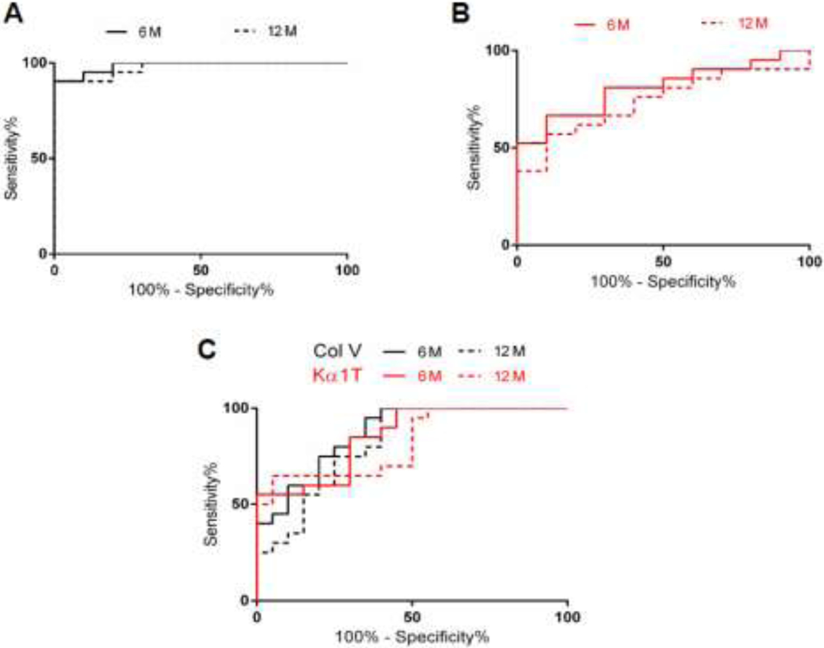
Background: Exosomes isolated from plasma of lung transplant recipients (LTxRs) with bronchiolitis obliterans syndrome (BOS) contain human leukocyte antigens and lung self-antigens (SAgs), K-alpha 1 tubulin (Kα1T) and collagen type V (Col-V). The aim was to determine the use of circulating exosomes with lung SAgs as a biomarker for BOS.

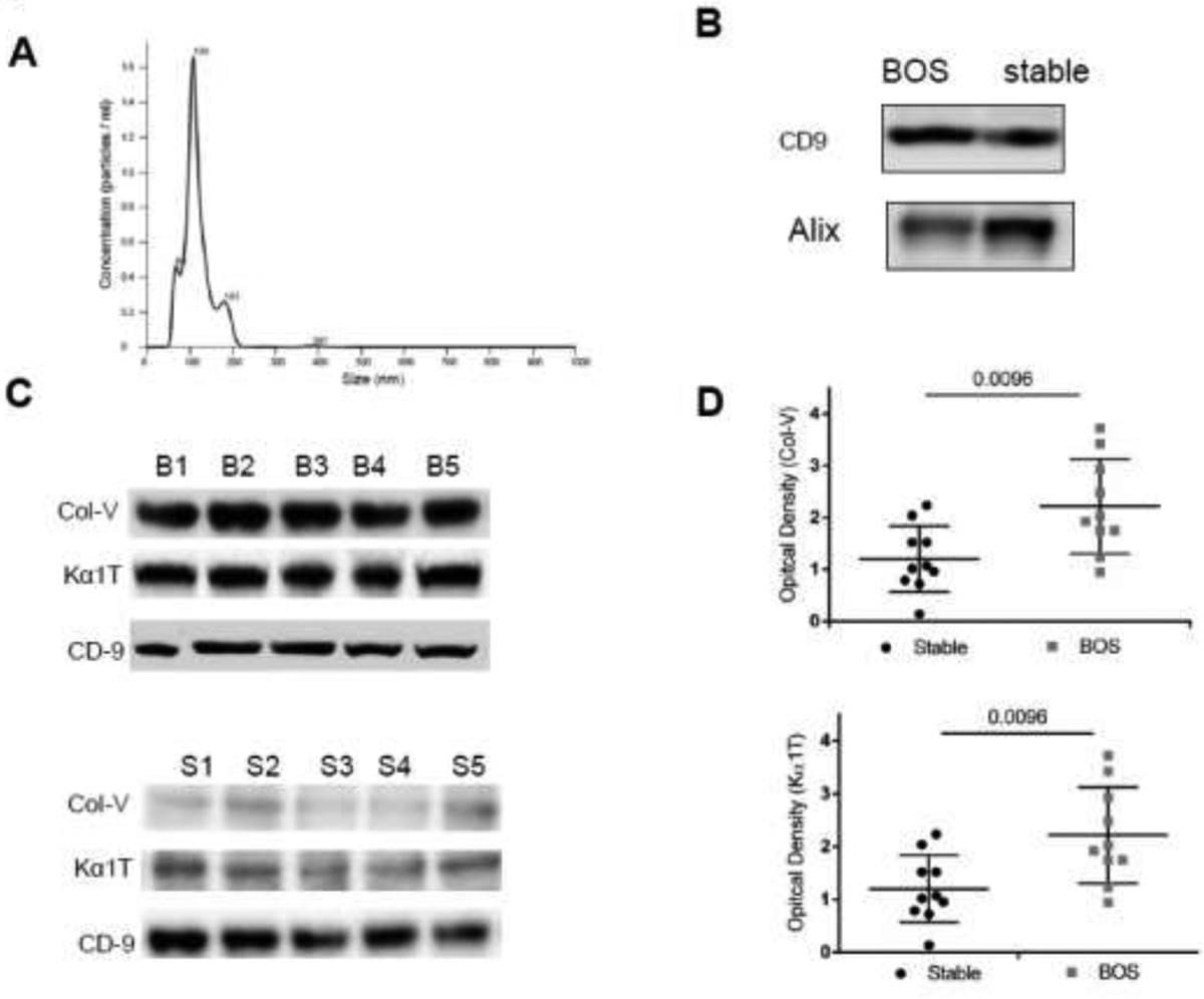
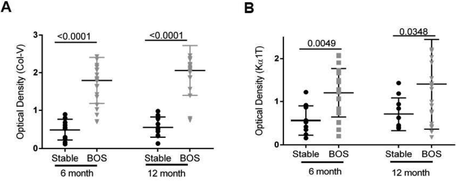
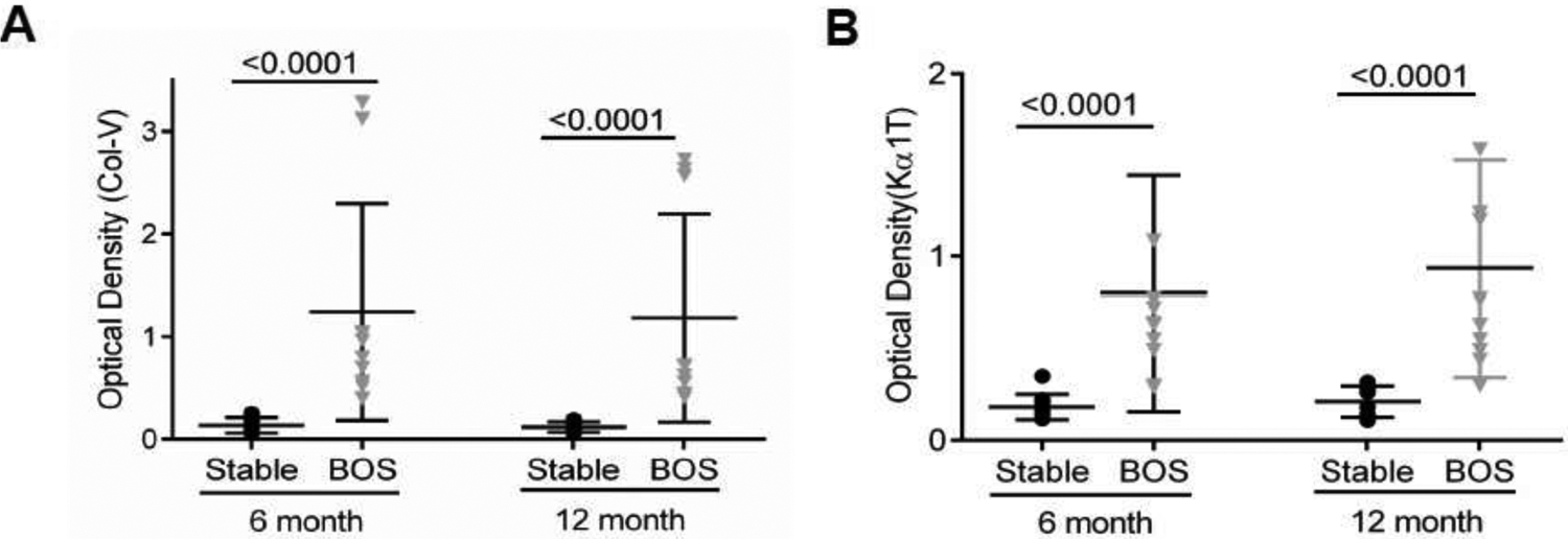
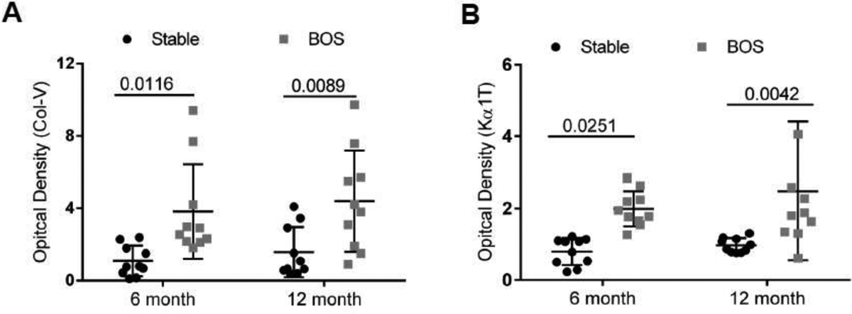
Methods: Circulating exosomes were isolated retrospectively from plasma from LTxRs at diagnosis of BOS and at 6 and 12 months before the diagnosis (n = 41) and from stable time-matched controls (n = 30) at 2 transplant centers by ultracentrifugation. Exosomes were validated using Nanosight, and lung SAgs (Kα1T and Col-V) were detected by immunoblot and semiquantitated using ImageJ software.

Results: Circulating exosomes from BOS and stable LTxRs demonstrated 61- to 181-nm vesicles with markers Alix and CD9. Exosomes from LTxRs with BOS (n = 21) showed increased levels of lung SAgs compared with stable (n = 10). A validation study using 2 separate cohorts of LTxRs with BOS and stable time-matched controls from 2 centers also demonstrated significantly increased lung SAgs-containing exosomes at 6 and 12 months before BOS.

Conclusions: Circulating exosomes isolated from LTxRs with BOS demonstrated increased levels of lung SAgs (Kα1T and Col-V) 12 months before the diagnosis (100% specificity and 90% sensitivity), indicating that circulating exosomes with lung SAgs can be used as a non-invasive biomarker for identifying LTxRs at risk for BOS.

Keywords: biomarker; chronic lung allograft dysfunction; circulating exosomes; human lung transplant; lung self-antigens.


Conflict of interest statement

Disclosure Statement:

The authors have no conflict of interest to disclose.

Figures

Figure 1:
Figure 1:
(A) Exosomes were isolated from plasma by ultracentrifugation, diluted in PBS and subjected to NanosightNS300 analysis and results demonstrated that the extracellular vesicles has size of 60–200 nM. Similar results were also obtained using the modified kit method. (B) Western blot analysis showed presence of exosomes markers CD9, Alix in exosomes isolated from plasma of stable and bronchiolitis obliterans syndrome (BOS) patients. (C) Exosomes isolated from plasma from a different center (UW) at the time of BOS diagnosis contain Collagen V (Col-V) and Kα1 Tubulin (Kα1T): Exosomes were isolated from plasma of lung transplant recipients (LTxRs) from University of Washington (different center) diagnosed with BOS (n=10) and stable (time match control) (n=10) and subjected to western blot analysis for the detection of lung self-antigens. Western blot analysis of representative 5 BOS and 5 stable showed: increased amount of Col-V and Kα1T in exosomes derived from BOS LTxRs but not in stable. Representation depicts 5 out of 10 LTxRs from BOS (top) and stable (bottom). (D) Semi quantification by densitometry showed significant increase in optical density for Col-V (top) and Kα1T (bottom) in bronchiolitis obliterans syndrome (BOS) lung transplant recipients when compared with stable.
Figure 2:
Figure 2:
Analysis of exosomes isolated from circulation of 21 BOS LTxR and 10 time matched stable LTxR (Discovery Cohort) (A). Semi-quantification by densitometry revealed significant increase in Col-V optical density in comparison to stable, 6 month (1.79±0.59 vs 0.49±0.27, p<0.0001) and 12 month (2.06±0.65 vs 0.56±0.26, p<0.0001). (B) Densitometry analysis showed significantly increased Kα1T optical density when compared with stable, 6 month (1.20±0.55 vs 0.56±0.34, p=0.0049) and 12 month (1.41±1.02 vs 0.71±0.37, p=0.0348). Western blot data is presented in Supplementary figure S1.
Figure 3:
Figure 3:
.Analysis of circulating exosomes for lung SAgs from a different LTxRs center (WUSM) (Validation cohort) consisting of 10 with BOS and 10 stable/control demonstrating increased levels of lung SAgs containing exosomes 6 and 12 months prior to clinical diagnosis of BOS. Densitometry analysis demonstrate that exosomes isolated from BOS patients has increased lung self-antigens, (Optical Density, Collagen V (Col-V) (A), 6 month 1.24±1.06 vs 0.13±0.07, p<0.0001; 12 months 1.18±1.02 vs .0.12±0.05, p<0.0001 (B) (Optical density Kα1 Tubulin (Kα1T) 6 months 0.80±0.64 vs 0.18±0.07, p<0.0001) and; 12 months 0.94±0.59 vs 0.21±0.09, p<0.0001). Western blot results are presented in Supplementary figure S2.
Figure 4:
Figure 4:
Analysis of circulating exosomes for lung SAgs from a different LTxRs center ( UW) (Validation cohort) consisting of 10 with BOS and 10 stable/control demonstrating increased levels of lung SAgs containing exosomes 6 and 12 months prior to clinical diagnosis of BOS. Exosomes isolated from plasma of BOS had significantly increased levels of lung self-antigens in comparison to stable. Semi quantification analysis of optical density of Collagen V (Col-V). (A) (6 month: 3.8±2.6 vs 1.09±0.84; p=0.0116, 12 month: 4.39±2.79 vs 1.57±1.39 p=0.0089), Kα1 Tubulin (Kα1T). (B) (6 month: 2.00±0.49 vs 0.080±0.37; p=0.0251, 12 month: 2.48±1.92 vs 0.98±0.19 p=0.0042. Western blot results are presented in Supplementary figure S3.
Figure 5.
Figure 5.
(A) Receiver operating curve (ROC) were calculated for circulating exosomes with lung self-antigen (SAg), Collagen V (Col-V), at two time points (6 and 12 months) in discovery cohort to determine optimum threshold values. Col-V levels at 6 months (—) had an area under curve (AUC) AUC=0.99 and 12 month (- - -) (AUC=0.98). (B) ROC was calculated for circulating exosomes with lung SAg, Kα1 Tubulin (Kα1T), at two time points (6 and 12 months) in discovery cohort to determine optimum threshold values. Kα1T levels had an AUC at 6 months (0.81) and 12 month (0.74). (C) Validation Cohort; ROC were calculated for circulating exosomes with lung SAgs at two time points (6 and 12 months) for combined validation cohorts from both centers (Washington Univ. School of Medicine and Univ. Washington-Seattle). Validation cohort revealed that Col-V levels at 6 months had AUC=0.87 and at 12 month (AUC=0.82) (black). Kα1T levels at 6 months had an AUC=0.85 and 12 months (AUC=0.82) respectively (Red).

References

    1. Estenne M, and Hertz MI. Bronchiolitis obliterans after human lung transplantation. Am J Resp Crit Care Med 2003;166 440–4. - PubMed
    1. Chambers DC, Yusen RD, Cherikh WS, et al. The Registry of the International Society for Heart and Lung Transplantation: Thirty-fourth Adult Lung And Heart-Lung Transplantation Report-2017; Focus Theme: Allograft ischemic time. J Heart Lung Transplant 2017;36:1047–59. - PubMed
    1. Pakhale SS, Hadjiliadis D, Howell DN, et al. Upper lobe fibrosis: a novel manifestation of chronic allograft dysfunction in lung transplantation. J Heart Lung Transplant 2005;24:1260–8. - PubMed
    1. Sato M, Waddell TK, Wagnetz U, et al. Restrictive allograft syndrome (RAS): a novel form of chronic lung allograft dysfunction. J Heart Lung Transplant 2011;30:735–42. - PubMed
    1. Burke CM, Theodore J, Dawkins KD, et al. Post-transplant obliterative bronchiolitis and other late lung sequelae in human heart-lung transplantation. Chest 1984;86:824–9. - PubMed

Publication types