. 2020 Jul 2:2020:3707904.
doi: 10.1155/2020/3707904. eCollection 2020.

Antidiabetic Effects of Arginyl-Fructosyl-Glucose, a Nonsaponin Fraction from Ginseng Processing in Streptozotocin-Induced Type 2 Diabetic Mice through Regulating the PI3K/AKT/GSK-3 β and Bcl-2/Bax Signaling Pathways

Affiliations

Antidiabetic Effects of Arginyl-Fructosyl-Glucose, a Nonsaponin Fraction from Ginseng Processing in Streptozotocin-Induced Type 2 Diabetic Mice through Regulating the PI3K/AKT/GSK-3 β and Bcl-2/Bax Signaling Pathways

Xinglong Liu et al. Evid Based Complement Alternat Med. .

Abstract

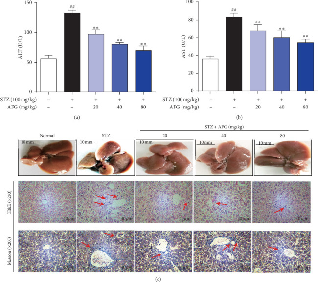
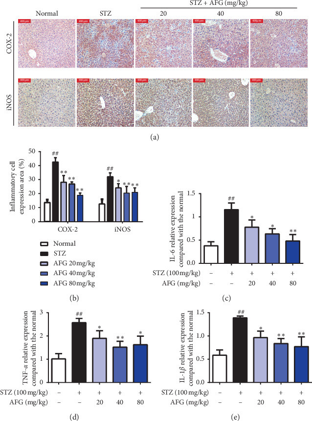
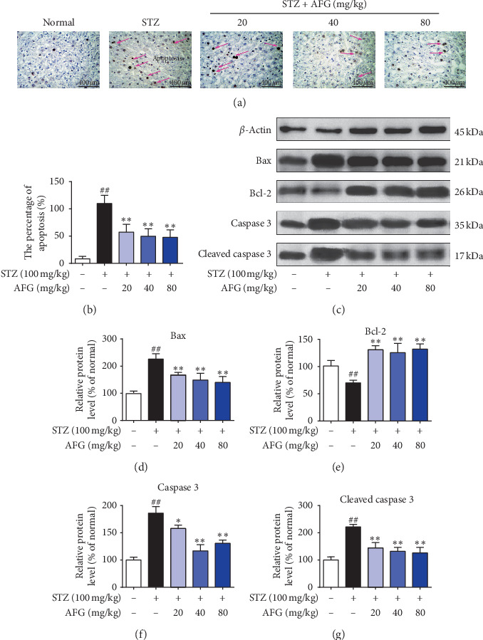
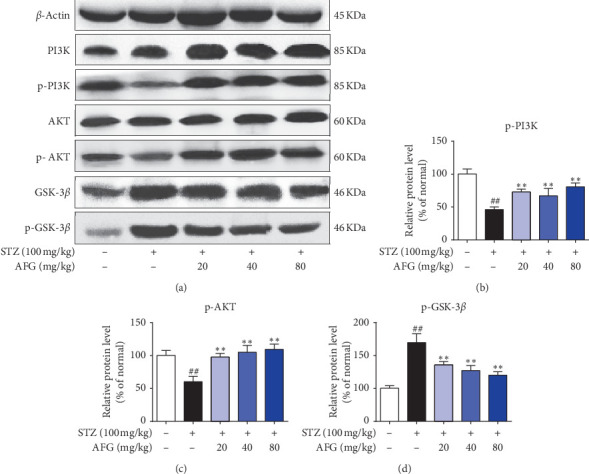
Streptozotocin- (STZ-) induced type 2 diabetes mellitus (T2DM) caused insulin secretion disorder and hyperglycemia, further causing tissue and organ damage. In recent years, studies on ginseng (Panax ginseng C. A. Meyer) and its saponins (Ginsenosides) have proved to possess antidiabetic pharmacological activities, but the mechanism of nonsaponins on STZ-induced T2DM is still unclear. Arginyl-fructosyl-glucose (AFG) is a representative nonsaponin component produced in the processing of red ginseng. The present study was designed to assess the possible healing consequence of AFG on STZ-induced T2DM in mice and also to explore its fundamental molecular contrivances. T2DM-related indexes, fasting blood glucose levels, and body weight, histological changes, biochemical considerations, biomarkers, the mRNA countenance intensities of inflammatory facts, and variations in correlated protein manifestation in adipose tissue and liver tissue were calculated. Consequences specified that AFG usage successfully amends STZ-induced insulin conflict and liver grievance in T2DM. Systematically, AFG action diminished STZ-induced oxidative stress and inflammatory responses in the liver. In addition, we demonstrated that AFG also attenuates apoptosis and insulin secretion disorders in T2DM by adjusting the PI3K/AKT/GSK3β signaling pathway. At the end, these discoveries recommend that AFG averts the development of T2DM through numerous types of machinery and proposes that AFG can also be used in order to treat T2DM in the future.


Conflict of interest statement

The authors declare no conflicts of interest.

Figures

Figure 1
Figure 1
AFG improves the blood glucose index of T2DM. (a) the Structure of Arginyl-fructosyl-glucose (AFG); (b) Fasting blood glucose (FBG) levels in different doses mice at different times; (c) Trend line of oral glucose tolerance test (OGTT) in diabetic mice (DM). Data are exhibitedas the mean ± S.D. (n = 12). #P < 0.05, ##P < 0.01, as equated with normal group; P < 0.05, ∗∗P < 0.01, as equated with STZ group.
Figure 2
Figure 2
Protective effect of AFG treatment on STZ-induced liver injury in T2DM mice. (a) Alanine aminotransferase (ALT) content in mice liver; (b) Aspartate aminotransferase (AST) content; (c) H&E and Masson staining of liver sections, magnification: ×400. Data explored as the mean ± S.D. (n = 12). #P < 0.05, ##P < 0.01, as equated with normal group; P < 0.05, ∗∗P < 0.01, as equated with STZ.
Figure 3
Figure 3
AFG mitigates oxidative stress and lipid metabolism disorder in STZ-induced T2DM. (a) Liver GSH activities; (b) Lipid peroxidation MDA; (c) Antioxidant enzyme SOD; (d) Liver GSH/GSSH level; (e–h) Effect of AFG on the expression of TC, TG, LDL-C and HDL-C in liver. Data presented as the mean ± S.D. (n = 12). #P < 0.05, ##P < 0.01, as equated with normal group; P < 0.05, ∗∗P < 0.01, as equated with STZ group.
Figure 4
Figure 4
Effects of AFG on the expression of inflammatory factors in liver tissue. (a, b) Analysis of iNOS and COX-2 expression in liver tissue. (c–e) Relative mRNA levels of TNF-α, IL-1β, and IL-6 in liver tissue. Data presented as the mean ± S.D. (n = 12). #P < 0.05, ##P < 0.01, as equated with the normal group; P < 0.05, ∗∗P < 0.01, as equated with the STZ group.
Figure 5
Figure 5
Effects of AFG antiapoptotic in STZ-induced liver tissue. (a, b) Evaluation of TUNEL staining and the percentage of apoptosis. (c) Determination of Bax, Bcl-2, caspase-3, Cleaved caspase-3 proteins expression, and β-actin as a reference. (d–g) Analysis of relative protein expression by integrated optical density. Data presented as the mean ± S.D. (n = 12). #P < 0.05, ##P < 0.01, as equated with the normal group; P < 0.05, ∗∗P < 0.01, as equated with the STZ group.
Figure 6
Figure 6
AFG regulates glycogen metabolism and ameliorates hepatocyte apoptosis in diabetic mice by adjusting the liver PI3K/AKT/GSK3β signaling pathway. (a) Possessions of AFG on the manifestation of PI3K/AKT/GSK3β in the liver tissues were explored by western blot exploration. (b–d) Densitometric examination of western blot. Data are expressed as the mean ± S.D. (n = 10). #P < 0.05, ##P < 0.01, as equated with the normal group; P < 0.05, ∗∗P < 0.01, as equated with the STZ group.
Figure 7
Figure 7
AFG improves STZ-induced changes in peritoneal fat and liver factors in T2DM mice. (a, c, e) Effects of AFG on the expression of SREBP-1, FAS, and MCP-1 in the peritoneal fat were measured by RT-PCR. (b, d) Special effects of AFG on the manifestation of IDE and HNF-4α in the liver tissue were measured by RT-PCR. Data are conveyed as the mean ± S.D. (n = 12). #P < 0.05, ##P < 0.01, as equated with the normal group; P < 0.05, ∗∗P < 0.01, as equated with the STZ group.

References

    1. Mehrabian S., Raycheva M., Gateva A., et al. Cognitive dysfunction profile and arterial stiffness in type 2 diabetes. Journal of the Neurological Sciences. 2012;322(1-2):152–156. doi: 10.1016/j.jns.2012.07.046. - DOI - PubMed
    1. Pang B., Yu X.-T., Zhou Q., et al. Innovative thoughts on treating diabetes from the perspective of traditional Chinese medicine. Evidence-Based Complementary and Alternative Medicine. 2015;2015:12. doi: 10.1155/2015/905432.905432 - DOI - PMC - PubMed
    1. Kahn R. Follow-up report on the diagnosis of diabetes mellitus: the expert committee on the diagnosis and classifications of diabetes mellitus. Diabetes Care. 2003;26:3160–3167. - PubMed
    1. Hosseini S. E., Tavakoli F., Karami M. Medicinal plants in the treatment of diabetes mellitus. Clinical Excellence. 2014;2:64–89.
    1. Hameed I., Masoodi S. R., Mir S. A., Nabi M., Ghazanfar K., Ganai B. A. Type 2 diabetes mellitus: from a metabolic disorder to an inflammatory condition. World Journal of Diabetes. 2015;6(4):598–612. doi: 10.4239/wjd.v6.i4.598. - DOI - PMC - PubMed